Footprint Identification Technology (FIT): A Comprehensive Guide to Implementation in Modern Biomedical Research
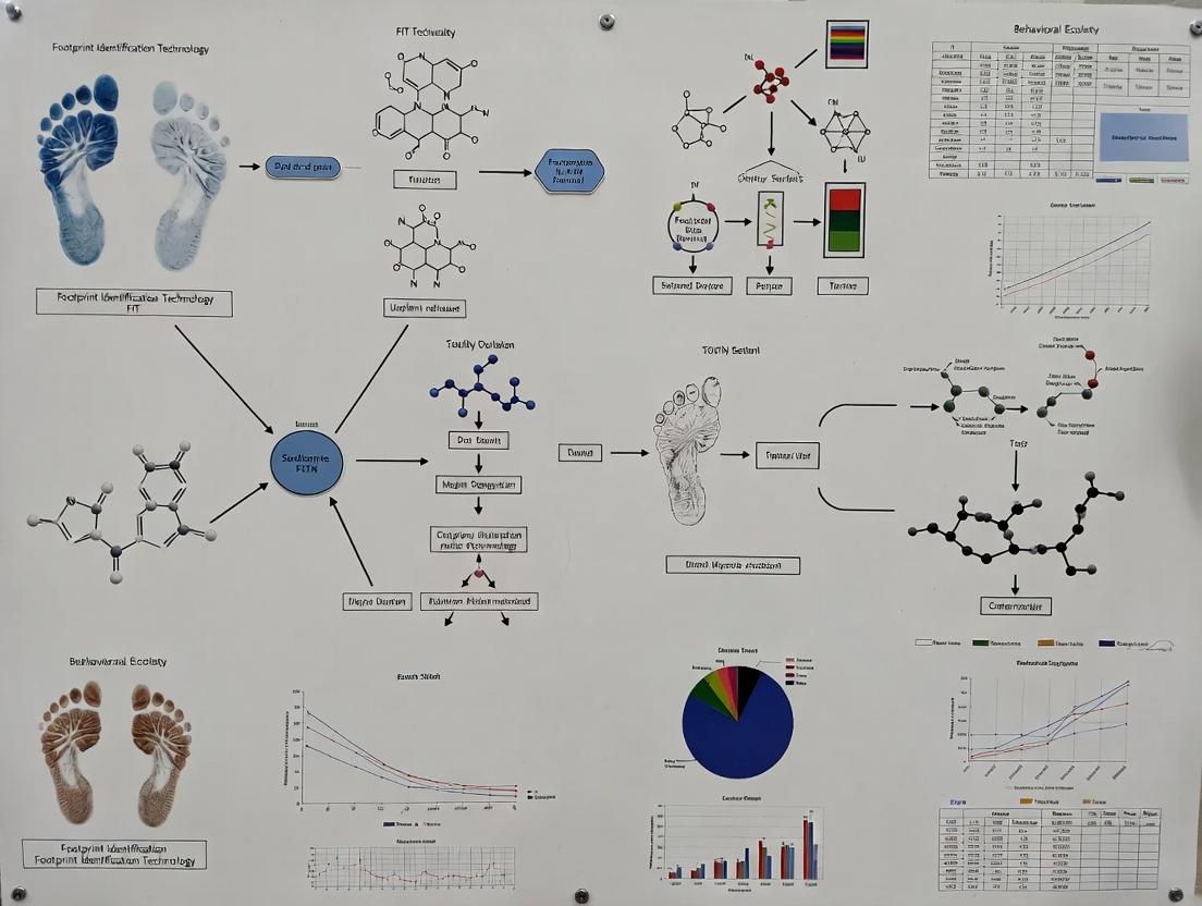
This article provides a comprehensive overview of Footprint Identification Technology (FIT) for researchers and drug development professionals.
Footprint Identification Technology (FIT): A Comprehensive Guide to Implementation in Modern Biomedical Research
Abstract
This article provides a comprehensive overview of Footprint Identification Technology (FIT) for researchers and drug development professionals. It explores the foundational principles of FIT as a high-resolution tool for mapping protein-DNA interactions and transcriptional regulation. The content details methodological workflows for chromatin preparation, library construction, sequencing, and data analysis, alongside practical applications in enhancer discovery and compound mechanism-of-action studies. It addresses common experimental and bioinformatic troubleshooting challenges and offers optimization strategies. Finally, the article validates FIT against established techniques like ChIP-seq and ATAC-seq, evaluating its sensitivity, specificity, and unique advantages to guide informed technology selection for epigenetic and transcriptional research.
What is Footprint Identification Technology? Core Principles and Research Applications
Historical Context and Evolution of FIT
Footprint Identification Technology (FIT), in a molecular biology context, traditionally refers to methods used to identify protein-binding sites on DNA, known as footprints. The core principle, established in the late 1970s, relies on the protection of DNA from cleavage or modification by a bound protein. The advent of high-throughput sequencing (HTS) has transformed FIT from a low-throughput, gel-based assay to a genome-wide discovery tool.
Table 1: Evolution of Footprinting Techniques
| Technique | Era | Principle | Throughput | Key Limitation |
|---|---|---|---|---|
| DNase I Footprinting | 1970s-2000s | DNase I cleaves exposed DNA; bound protein protects site. | Low (single locus) | Requires prior knowledge of binding region. |
| In Vivo Footprinting | 1990s-2010s | Uses chemical agents (e.g., DMS) in living cells to assess protein accessibility. | Low to Medium | Complex analysis, often limited to known sites. |
| Digital Genomic Footprinting (DGF) | 2010s-Present | DNase I or Tn5 cleavage coupled with HTS (DNase-seq, ATAC-seq). | High (genome-wide) | Identifies footprints indirectly via cleavage patterns. |
| Protein-Specific Footprinting | 2010s-Present | Use of engineered nucleases (e.g., ChIP-exo, CUT&RUN, CUT&Tag). | High (genome-wide) | Provides direct, protein-specific binding site maps. |
Modern FIT Protocols: ATAC-seq as a Paradigm
Assay for Transposase-Accessible Chromatin with high-throughput sequencing (ATAC-seq) is a contemporary FIT method that identifies open chromatin regions and, via computational footprinting, infers transcription factor (TF) binding sites.
Protocol: ATAC-seq for Nucleosome and TF Footprint Mapping
I. Cell Preparation and Transposition
- Cell Lysis: Harvest 50,000-100,000 viable cells. Pellet and resuspend in cold lysis buffer (10 mM Tris-HCl pH 7.4, 10 mM NaCl, 3 mM MgCl2, 0.1% IGEPAL CA-630). Incubate on ice for 3 minutes.
- Nuclei Isolation: Pellet nuclei immediately at 500 x g for 10 minutes at 4°C. Resuspend pellet in transposition mix.
- Tagmentation: Prepare a 50 µL reaction containing 25 µL 2x TD Buffer, 2.5 µL Tn5 Transposase (Illumina), 16.5 µL PBS, 0.5 µL 1% Digitonin, 0.5 µL 10% Tween-20, and 5 µL of nuclei suspension. Incubate at 37°C for 30 minutes in a thermomixer with shaking.
- DNA Clean-up: Immediately purify tagmented DNA using a SPRI bead-based cleanup system (e.g., Zymo DNA Clean & Concentrator-5). Elute in 21 µL Elution Buffer.
II. Library Amplification and Sequencing
- PCR Amplification: Perform a 50 µL PCR reaction using 2x KAPA HiFi HotStart ReadyMix and 1.25 µM of indexed forward and reverse primers. Use a cycling protocol with 5-10 cycles, determined by a 5 µL qPCR side reaction to prevent over-amplification.
- Library Clean-up: Purify the final library using a double-sided SPRI bead clean-up (e.g., 0.5x followed by 1.2x ratios) to select fragments primarily between 150-800 bp. Quantify via qPCR or bioanalyzer.
- Sequencing: Sequence on an Illumina platform, typically 75 bp paired-end, aiming for 50-100 million reads per sample.
Visualization of Workflows and Pathways
Title: ATAC-seq Experimental Workflow
Title: Logic of TF Footprinting from ATAC-seq Data
The Scientist's Toolkit: Key Reagents for Modern FIT
Table 2: Essential Research Reagents for ATAC-seq-based FIT
| Item | Function in Protocol | Example/Note |
|---|---|---|
| Tn5 Transposase | Engineered transposase that simultaneously fragments ("tagments") DNA and adds sequencing adapters. Core enzyme of ATAC-seq. | Illumina Tagment DNA TDE1 Enzyme, or homemade loaded enzyme. |
| Digitonin | Mild detergent used to permeabilize the nuclear membrane, allowing Tn5 access to chromatin while maintaining nuclear integrity. | Critical for optimizing in-nucleus tagmentation efficiency. |
| SPRI Magnetic Beads | Size-selective solid-phase reversible immobilization beads for post-tagmentation clean-up and PCR product size selection. | Zymo, Beckman Coulter, or equivalent. Key for removing large fragments (>800 bp). |
| High-Fidelity PCR Mix | Robust polymerase for minimal-bias amplification of the tagmented library. Essential for maintaining complexity. | KAPA HiFi HotStart ReadyMix, NEB Next High-Fidelity. |
| Dual-Indexed PCR Primers | Unique barcoded primers for multiplexing samples during sequencing. Allow pooling of multiple libraries. | Illumina Nextera-style indices, IDT for Illumina. |
| Cell Viability Stain | Critical for selecting only live, intact cells/nuclei for input, as dead cells contribute high background. | Trypan Blue, DAPI, or Propidium Iodide for FACS. |
| Nuclei Counter | Accurate quantification of nuclei concentration is essential for optimizing tagmentation reaction input. | Automated cell counter or hemocytometer. |
Article Context: Footprint Identification Technology (FIT) Implementation Research
Footprint Identification Technology (FIT) is a cornerstone methodology in functional genomics for mapping protein-DNA interactions in vitro and in vivo. The core biochemical principle underpinning FIT is the differential sensitivity of DNA to nucleases like DNase I or Micrococcal Nuclease (MNase) when bound by regulatory proteins. Protein-bound DNA is protected from cleavage, creating a "footprint" of inaccessibility. This document details the application of this principle in modern research, providing protocols and resources for its implementation.
DNase I and MNase are endonucleases used to probe chromatin architecture and transcription factor occupancy. DNase I preferentially cleaves nucleosome-depleted, accessible regions, while MNase preferentially digests linker DNA between nucleosomes. Bound proteins, such as transcription factors or nucleosomes, sterically hinder enzyme access, resulting in reduced cleavage (a "protected" footprint) flanked by regions of enhanced cleavage due to protein-induced DNA distortion. FIT leverages high-throughput sequencing of these cleavage patterns (DNase-seq, MNase-seq) to identify protected footprints at single-nucleotide resolution, cataloging functional regulatory elements genome-wide.
Table 1: Comparative Properties of DNase I and MNase in Footprinting Assays
| Property | DNase I | MNase (Micrococcal Nuclease) |
|---|---|---|
| Primary Application in FIT | Mapping hypersensitive sites & transcription factor footprints in open chromatin. | Mapping nucleosome positions & boundaries; finer resolution of protein complexes. |
| Optimal Digestion Temperature | 37°C | 25-37°C (often 25°C for controlled digestion) |
| Key Cofactor Requirement | Ca²⁺, Mg²⁺ / Mn²⁺ | Ca²⁺ |
| Typical Digestion Time | 1-15 minutes | 5-20 minutes |
| Typical Enzyme Concentration Range | 0.1 - 5 units/µL (highly sample-dependent) | 0.01 - 0.5 units/µL (highly sample-dependent) |
| Primary Cleavage Product | Double-stranded breaks, blunt ends or 5'-P overhangs. | Single-stranded nicks leading to double-strand breaks; produces mononucleosomes. |
| Readout | Sequencing of cleavage ends (DNase-seq). | Sequencing of protected fragments (MNase-seq). |
| Primary Challenge | Determining optimal digestion concentration for footprint resolution. | Over-digestion leading to nucleosome displacement. |
Table 2: Typical FIT Workflow Metrics from Recent Studies (2023-2024)
| Workflow Step | Typical Yield/Output | Quality Control Checkpoint |
|---|---|---|
| Nuclei Isolation | 1-10 million nuclei per condition. | Trypan Blue viability >85%, intact nuclei via microscopy. |
| Titration Digestion | Varies; aim for >80% sub-nucleosomal fragments (DNase) or ~70-80% mononucleosomes (MNase). | Agarose gel electrophoresis "ladder" pattern. |
| Library Prep (Post-digestion) | Final library concentration: 5-30 nM. | Bioanalyzer/TapeStation profile: peak ~200-500 bp. |
| Sequencing | 20-50 million paired-end reads per sample (human/mouse). | >70% of reads uniquely mapped, low PCR duplicate rate. |
| Bioinformatic Footprint Calling | Identifies 50,000-200,000 footprints per cell type. | Correlation with known transcription factor motifs (e.g., ENCODE), reproducibility between replicates. |
Experimental Protocols
Protocol 1: DNase I Hypersensitivity & Footprinting on Isolated Nuclei
Objective: To generate a genome-wide map of DNase I cleavage sites and protected footprints from mammalian tissue culture cells.
Materials: See "Research Reagent Solutions" below.
Method:
- Nuclei Isolation: Harvest 5-10 million cells. Wash with cold PBS. Resuspend in 1 mL of cold Lysis Buffer (10 mM Tris-Cl pH 8.0, 10 mM NaCl, 3 mM MgCl2, 0.1% IGEPAL CA-630). Incubate on ice for 10 min. Pellet nuclei (500 x g, 5 min, 4°C). Wash once with 1 mL of cold Dignam Buffer C (20 mM HEPES pH 7.9, 25% glycerol, 1.5 mM MgCl2, 0.42 M NaCl, 0.2 mM EDTA). Resuspend nuclei in 100 µL of cold DNase I Digestion Buffer (15 mM Tris-Cl pH 8.0, 60 mM KCl, 15 mM NaCl, 1 mM CaCl2, 0.34 M sucrose).
- Titration & Digestion: Aliquot nuclei into 5 tubes. Add increasing concentrations of DNase I (e.g., 0, 0.5, 1, 2, 4 units) in a 50 µL reaction. Incubate at 37°C for exactly 3 minutes.
- Reaction Termination: Immediately add 100 µL of Stop Buffer (50 mM Tris-Cl pH 8.0, 100 mM NaCl, 0.1% SDS, 100 mM EDTA, 1 mM EGTA, 0.34 M sucrose). Add 2 µL of Proteinase K (20 mg/mL). Incubate at 55°C for 2 hours.
- DNA Purification: Add RNase A, incubate 30 min at 37°C. Purify DNA using phenol:chloroform:isoamyl alcohol extraction and ethanol precipitation. Resuspend in TE buffer.
- Size Selection & Library Prep: Run digested DNA on a 1.5% agarose gel. Excise the smear corresponding to 100-500 bp fragments (sub-nucleosomal). Gel-purify DNA. Use this size-selected DNA as input for a standard Illumina sequencing library preparation kit, incorporating steps to repair ends, add adapters, and PCR amplify.
- Sequencing & Analysis: Sequence on an Illumina platform (PE 50-100 bp). Map reads to reference genome. Identify cleavage hotspots (DHSs) and protected footprints using algorithms like Centipede, Wellington, or HINT.
Protocol 2: MNase Digestion for Nucleosome & Factor Footprinting
Objective: To map nucleosome positions and fine-scale protein-DNA interactions using MNase.
Materials: See "Research Reagent Solutions" below.
Method:
- Nuclei Preparation: Prepare nuclei as in Protocol 1, Step 1, but resuspend final pellet in 100 µL of MNase Digestion Buffer (10 mM Tris-Cl pH 7.5, 15 mM NaCl, 60 mM KCl, 0.34 M Sucrose, 0.15 mM spermine, 0.5 mM spermidine, 1 mM CaCl2).
- Titration Digestion: Aliquot nuclei. Add increasing concentrations of MNase (e.g., 0, 0.5, 2, 8, 32 units/mL) in a 50 µL reaction. Incubate at 25°C for 10 minutes.
- Reaction Termination: Add 50 µL of Stop Solution (100 mM EDTA, 10 mM EGTA, 1% SDS). Add Proteinase K (2 µL of 20 mg/mL). Incubate at 55°C overnight.
- DNA Purification & Analysis: Purify DNA as in Protocol 1, Step 4. Analyze 1 µg on a 1.5% agarose gel to visualize the mononucleosome ladder (~150 bp). Optimize digestion to yield ~80% mononucleosomes.
- Library Preparation for Protected Fragments: For nucleosome positioning, gel-purify mononucleosomal DNA (~140-160 bp). For fine-resolution footprinting, gel-purify shorter fragments (50-120 bp) corresponding to sub-nucleosomal, protein-protected DNA. Proceed with Illumina library prep.
- Sequencing & Analysis: Sequence and map reads. For nucleosome mapping, use peak-calling algorithms. For footprints, use MNase-based footprinting algorithms.
Visualizations
Title: FIT Workflow: From Cells to Footprints
Title: Biochemical Principle of Nuclease Footprinting
The Scientist's Toolkit
Table 3: Essential Research Reagent Solutions for FIT
| Item | Function in FIT | Key Considerations |
|---|---|---|
| DNase I (RNase-free) | Enzyme for probing open chromatin and TF footprints. | Purchase high-purity, recombinant grade. Aliquot and store at -20°C. Critical to titrate for each cell type. |
| Micrococcal Nuclease (MNase) | Enzyme for nucleosome mapping and fine-resolution footprinting. | S. aureus origin. Activity is highly dependent on Ca²⁺ concentration. |
| IGEPAL CA-630 (NP-40) | Non-ionic detergent for cell membrane lysis during nuclei isolation. | Less harsh than SDS, preserves nuclear membrane integrity. |
| Spermidine & Spermine | Polyamines added to MNase buffers. | Stabilize chromatin structure during digestion, preventing aggregation. |
| Protease Inhibitor Cocktail (PIC) | Added to all buffers during nuclei prep. | Prevents proteolytic degradation of DNA-binding proteins of interest. |
| Size Selection Beads | Magnetic beads (e.g., SPRI/AMPure) for DNA cleanup and size selection. | Critical for isolating sub-nucleosomal or mononucleosomal DNA fragments post-digestion. |
| Illumina-Compatible Library Prep Kit | For preparing sequencing libraries from low-input, fragmented DNA. | Choose kits optimized for FFPE or ChIP-seq samples, as they handle short, damaged DNA well. |
| High-Sensitivity DNA Assay | Fluorometric assay (e.g., Qubit) for accurate quantification of diluted, small DNA fragments. | More accurate than absorbance (Nanodrop) for fragmented DNA post-digestion. |
Application Notes
Within the broader thesis on Footprint Identification Technology (FIT) implementation research, the generation of nucleotide-resolution TFBS maps is the foundational analytical output. These maps are not merely lists of binding loci; they represent comprehensive, high-definition atlases of protein-DNA interactions across the genome. For researchers and drug development professionals, these maps are critical for elucidating transcriptional regulatory networks, identifying non-coding disease variants, and validating on-target/off-target effects of novel therapeutics.
The core principle of FIT-based methods (e.g., DNase-seq, ATAC-seq, and their derivatives) is the detection of protected "footprints" within regions of open chromatin, corresponding to the exact genomic coordinates where a transcription factor (TF) is bound. Modern implementations integrate this footprint signal with motif analysis, chromatin accessibility quantitation, and often, paired gene expression data to generate predictive and functional models of regulation.
Key Quantitative Benchmarks: Recent advancements have significantly improved the resolution and accuracy of footprinting. The following table summarizes performance metrics from contemporary studies (2023-2024) comparing different algorithms and experimental couplings.
Table 1: Performance Metrics of Modern FIT-Based Footprinting Methods (2023-2024)
| Method / Algorithm | Experimental Coupling | Resolution (bp) | Validation Accuracy (AUC) | Key Advantage |
|---|---|---|---|---|
| Protein-informed Footprinting | ATAC-seq + TF ChIP-seq | 1-5 | 0.91-0.95 | Direct integration of protein binding data for training. |
| MILLIPEDE | High-depth DNase-seq | 4-8 | 0.88-0.93 | Models cleavage bias explicitly; high specificity. |
| HINT-ATAC | Standard ATAC-seq | 6-10 | 0.85-0.90 | Optimized for low-cell-number ATAC-seq data. |
| Binary Event Model (BEM) | DNase I or ATAC-seq | 1 (theoretical) | 0.82-0.87 | Focuses on single-nucleotide cleavage events. |
| ArchR | ArchR-linked ATAC-seq | 6-12 | 0.86-0.89 | Integrated single-cell multi-ome analysis. |
Experimental Protocols
Protocol 1: High-Resolution TFBS Mapping Using Protein-Informed Footprinting with ATAC-seq
Objective: To generate nucleotide-resolution TFBS maps by integrating ATAC-seq footprint signals with prior knowledge from TF-specific ChIP-seq data.
Materials: Fresh or frozen cell pellets (50k-100k cells), ATAC-seq kit (e.g., Illumina Tagmentase TDE1), SPRI beads, Qubit fluorometer, Bioanalyzer/TapeStation, PCR thermocycler, sequencing platform (e.g., Illumina NovaSeq).
Procedure:
- Cell Lysis & Tagmentation: Resuspend cells in ice-cold lysis buffer (10 mM Tris-HCl pH 7.4, 10 mM NaCl, 3 mM MgCl2, 0.1% IGEPAL CA-630). Incubate 3 min on ice. Pellet nuclei and immediately tagment DNA using the TDE1 enzyme (37°C, 30 min with shaking).
- DNA Purification: Clean up tagmented DNA using SPRI beads at a 1.8X ratio. Elute in 20 µL TE buffer.
- Library Amplification & Indexing: Amplify the purified DNA via PCR (12-15 cycles) using indexed primers compatible with your sequencer. Include a SYBR Green dye in a pilot reaction to determine the optimal cycle number before saturation.
- Library Purification & QC: Perform a double-sided SPRI bead cleanup (0.5X followed by 1.5X ratio) to remove primer dimers and large fragments. Quantify library concentration (Qubit) and profile fragment size distribution (Bioanalyzer High Sensitivity DNA chip). Aim for the characteristic ~200bp periodicity.
- Sequencing: Pool libraries and sequence on an Illumina platform. For footprinting, obtain high sequencing depth (>100 million paired-end 50-75bp reads per sample).
- Bioinformatic Analysis:
a. Preprocessing: Trim adapters (Cutadapt). Align reads to the reference genome (hg38/mm10) using a spliced-aware aligner (BWA-MEM, Bowtie2) with options to retain only properly paired, uniquely mapped, non-mitochondrial reads.
b. Footprint Calling: Process aligned BAM files to identify cleavage sites (5' ends of reads). Use the
TOBIASsuite: i.TOBIAS ATACorrect-- Corrects for Tn5 insertion sequence bias. ii.TOBIAS FootprintScores-- Calculates footprint scores per nucleotide using a sliding window. iii.TOBIAS BINDetect-- Integrates footprint scores with pre-defined TF motifs (from JASPAR) and optional ChIP-seq peak BED files to call bound/unbound sites. This is the "protein-informed" step. c. Output: The final output is a BED-like file with genomic coordinates, TF name, binding score, strand, and motif match, constituting the nucleotide-resolution TFBS map.
Protocol 2: In Silico Validation and Functional Annotation of TFBS Maps
Objective: To validate the accuracy of predicted TFBS and annotate them with potential target genes and disease associations.
Materials: Predicted TFBS map (BED file), reference genome, annotation files (e.g., GENCODE), disease SNP databases (GWAS Catalog, ClinVar), high-performance computing cluster.
Procedure:
- Validation via Overlap Analysis:
a. Download publicly available ChIP-seq peak data for relevant TFs from ENCODE or CistromeDB.
b. Use
bedtools intersectto calculate the percentage of predicted TFBS that overlap experimental ChIP-seq peaks (within a ±50bp window). A high overlap rate (>70%) indicates strong predictive accuracy. - Functional Gene Linking:
a. Annotate each TFBS with the nearest transcription start site (TSS) using
bedtools closest. b. For more accurate linking, use chromatin interaction data (e.g., promoter-capture Hi-C) if available for your cell type. Assign TFBS to genes based on significant chromatin loops. - Disease Variant Enrichment:
a. Download curated GWAS SNPs and their linked traits.
b. Use
bedtools intersectto identify TFBS that colocalize with GWAS SNPs. Perform an enrichment test (Fisher's exact test) to determine if specific traits are statistically overrepresented in your TFBS set. - Pathway Analysis:
a. Extract the list of target genes linked in Step 2.
b. Perform gene ontology (GO) and Kyoto Encyclopedia of Genes and Genomes (KEGG) pathway enrichment analysis using tools like
clusterProfileror Enrichr. Identify biological processes and pathways most regulated by the mapped TFs.
Title: Protein-Informed Footprinting Workflow
Title: Logic of Protein-Informed TFBS Detection
The Scientist's Toolkit
Table 2: Key Research Reagent Solutions for Nucleotide-Resolution TFBS Mapping
| Item | Function in Protocol | Example Product/Catalog |
|---|---|---|
| Tn5 Transposase | Enzyme that simultaneously fragments ("tagments") DNA and adds sequencing adapters in ATAC-seq. Essential for open chromatin profiling. | Illumina Tagmentase TDE1 (20034197) |
| SPRI Beads | Magnetic beads for size-selective purification and cleanup of DNA libraries. Critical for removing primers, dimers, and large fragments. | Beckman Coulter AMPure XP (A63881) |
| High-Sensitivity DNA Assay | Accurate quantification and size distribution analysis of final sequencing libraries prior to pooling. | Agilent High Sensitivity DNA Kit (5067-4626) |
| Indexed PCR Primers | Adds unique dual indexes (UDIs) to each library during amplification, enabling sample multiplexing in a single sequencing run. | Illumina IDT for Illumina UD Indexes (20027213) |
| Cell Lysis Buffer | Gently lyses cell membrane while leaving nuclei intact, a critical first step for clean ATAC-seq. | 10x Genomics Nuclei Buffer (2000207) or homemade (see protocol). |
| TF Motif Database | Curated collection of position weight matrices (PWMs) for known TFs, used for in silico motif scanning within footprint regions. | JASPAR (jaspar.genereg.net) |
| ChIP-seq Reference Data | Publicly available experimental TF binding data for training and validation of footprinting algorithms. | ENCODE Portal (encodeproject.org) |
Application Note: Utilizing FIT for Enhancer Validation and Network Inference
Footprint Identification Technology (FIT), leveraging assays like ATAC-seq and DNase-seq coupled with specialized computational pipelines, enables the genome-wide mapping of transcription factor (TF) binding events. This application note details its primary use in decoding transcriptional logic for therapeutic target discovery.
Table 1: Comparative Output of FIT-Enabled Assays
| Assay | Primary Output | Key Metric | Typical Resolution | Primary Application in Network Decoding |
|---|---|---|---|---|
| ATAC-seq | Open chromatin regions, nucleosome positions | Insertion site counts | ~100 bp | Identification of candidate CREs (enhancers, promoters) |
| DNase-seq | DNase I hypersensitive sites (DHS) | Cleavage frequency | ~150 bp | Delineation of broad regulatory regions |
| FIT Analysis | Protein-binding footprints within open chromatin | Footprint depth/score | 6-40 bp (exact TF binding site) | Inference of active TF binding events and identity |
Protocol 1: Integrated ATAC-seq and FIT Pipeline for TF Footprinting
Objective: To identify active cis-regulatory elements and bound transcription factors from mammalian cells.
Materials & Reagents:
- Nuclei Isolation Buffer: (10 mM Tris-Cl pH 7.4, 10 mM NaCl, 3 mM MgCl2, 0.1% IGEPAL CA-630). Gently lyses plasma membrane while preserving nuclear integrity.
- Tn5 Transposase (Tagmentase): Engineered hyperactive transposase pre-loaded with sequencing adapters (Nextera). Simultaneously fragments open chromatin and adds adapter sequences.
- Magnetic Size Selection Beads (SPRI): Paramagnetic beads for post-tagmentation DNA purification and size selection to enrich for nucleosome-free fragments.
- High-Fidelity PCR Mix: For limited-cycle PCR to amplify library fragments with unique dual indexing primers.
- Footprinting-Capable Software (e.g., HINT-ATAC, TOBIAS): Computational packages designed to detect statistically significant depletion of Tn5 insertion events, indicating protein protection.
Procedure:
- Cell Harvesting & Lysis: Pellet 50,000-100,000 viable cells. Resuspend in cold nuclei isolation buffer, incubate on ice, and pellet nuclei.
- Tagmentation: Resuspend nuclei in transposase reaction mix. Incubate at 37°C for 30 minutes. Immediately purify DNA using SPRI beads.
- Library Amplification: Perform PCR on purified DNA (5-12 cycles) using indexed primers. Purify final library with SPRI beads, selecting for fragments primarily below 700 bp.
- Sequencing: Perform paired-end sequencing (e.g., 2x50 bp) on an Illumina platform. Aim for ~50-100 million non-duplicate reads per sample for robust footprinting.
- Bioinformatic Analysis:
a. Preprocessing: Align reads to reference genome (e.g., hg38) using BWA or Bowtie2. Filter duplicates and mitochondrial reads.
b. Peak Calling: Call broad open chromatin regions using MACS2.
c. Footprint Detection: Run HINT-ATAC with the
-atacflag on aligned BAM files and peak regions. This identifies precise footprint locations. d. Motif Inference & TF Attribution: Annotate footprints using TOBIAS, which compares footprint scores against known TF motif databases (JASPAR, CIS-BP) to infer bound TFs.
The Scientist's Toolkit: Key Research Reagent Solutions
Table 2: Essential Reagents for FIT-Based Studies
| Item | Function | Example Product/Kit |
|---|---|---|
| Chromatin Accessibility Assay Kit | Standardized reagents for consistent nuclei preparation, tagmentation, and library prep. | Illumina ATAC-seq Kit, Nuclei Isolation Kit |
| Validated TF Antibodies | For ChIP-seq validation of specific TF binding events predicted by FIT. | CST, Abcam, Diagenode antibodies |
| TF Motif Database | Curated collection of position weight matrices (PWMs) for TF binding specificity. | JASPAR, CIS-BP, HOCOMOCO |
| Footprinting Software Suite | Integrated tools for alignment, peak calling, footprint detection, and TF annotation. | HINT-ATAC, TOBIAS, PIQ |
| CRISPR Activation/Interference (a/i) Systems | Functional validation of candidate CREs and TFs identified via FIT. | dCas9-VPR (activation), dCas9-KRAB (interference) |
Protocol 2: Constructing a Transcriptional Network from FIT-Derived Data
Objective: To integrate footprint data with transcriptomics to build a causal TF-to-target gene regulatory network.
Materials: FIT-derived TF binding list (from Protocol 1), matched RNA-seq data (from same cell type), gene annotation file (GTF), regulatory network software (e.g., GRNBoost2, SCENIC).
Procedure:
- Data Integration: Create a regulatory potential matrix. Associate each FIT-identified TF binding event with potential target genes (e.g., genes with a promoter or enhancer within ±500 kb of the footprint).
- Infer Regulatory Links: Using the co-expression data (RNA-seq) and the binding potential matrix, run GRNBoost2. This algorithm uses gradient boosting to infer robust, directional TF-to-target gene links.
- Network Pruning & Validation: Prune low-confidence links. Use the SCENIC pipeline to perform cis-regulatory motif enrichment analysis on the target genes for each TF, confirming the links are supported by both footprinting (binding) and motif evidence (specificity).
- Visualization & Analysis: Import the final adjacency list into Cytoscape. Identify network hubs (highly connected TFs), regulatory modules, and key target genes associated with disease pathways.
Diagrams
Title: FIT Analysis Workflow from Data to Network
Title: Core Transcriptional Regulatory Unit
Framing Context: This application note is developed as part of a thesis on the systematic implementation and validation of Footprint Identification Technology (FIT). It aims to provide a practical, data-driven comparison for researchers integrating high-specificity footprinting into chromatin and drug discovery pipelines.
FIT and general nuclease accessibility assays (e.g., DNase-seq, ATAC-seq) both probe DNA accessibility but differ fundamentally in resolution and information output.
Table 1: Assay Comparison - Specifications and Outputs
| Feature | General Nuclease Accessibility (ATAC-seq/DNase-seq) | Footprint Identification Technology (FIT) |
|---|---|---|
| Primary Objective | Map regions of open chromatin/genome-wide accessibility. | Identify precise protein-binding sites within accessible regions. |
| Nuclease/Agent | Transposase (ATAC) or DNase I (DNase-seq). | DNase I or micrococcal nuclease (MNase) at limited, titrated concentrations. |
| Key Readout | Reads clustered in open regions (peaks). | Depletions of reads at protein-bound sites within peaks (footprints). |
| Resolution | 100-500 bp open region. | Single-base pair resolution of protein-DNA interaction boundaries. |
| Informational Depth | Accessibility landscape. | Transcription factor (TF) identity (via footprint motif) and occupancy. |
| Typical Data Yield | ~50,000-150,000 accessible peaks per mammalian cell. | ~20,000-100,000 individual footprints within those peaks. |
| Drug Discovery Utility | Identify regulatory regions affected by treatment. | Directly map displacement or alteration of specific TF binding due to drug action. |
Table 2: Performance Metrics in a Model Study (K562 Cells)
| Metric | ATAC-seq (Standard) | FIT-DNase (from Thesis Data) |
|---|---|---|
| Total Peaks Called | 124,500 | N/A (analyzes peaks from accessibility assay) |
| Footprints Identified within Peaks | Not Applicable | 87,342 |
| Footprints with Significant TF Motif Match | Not Applicable | 68,901 (78.9%) |
| Signal-to-Noise Ratio (Footprint Depth) | N/A | 5.2:1 (protected vs. flanking cleavage) |
| Reproducibility (Pearson R between reps) | 0.98 (peak signal) | 0.93 (footprint call overlap) |
Detailed Experimental Protocols
Protocol A: FIT-DNase-seq for High-Resolution Footprinting
This protocol is optimized from the thesis implementation work.
I. Cell Preparation and Nuclei Isolation
- Harvest 1x10^6 cells, wash with cold PBS.
- Lyse in 5 mL of Hypotonic Lysis Buffer (10 mM Tris-Cl pH 7.4, 10 mM NaCl, 3 mM MgCl2, 0.1% IGEPAL CA-630, 1% protease inhibitor) on ice for 10 min.
- Pellet nuclei (500 x g, 5 min, 4°C). Wash once with 1 mL of Digestion Buffer (DB: 10 mM Tris-Cl pH 7.4, 10 mM NaCl, 3 mM MgCl2, 10% glycerol).
- Resuspend nuclei in DB to a concentration of ~1x10^6 nuclei/100 µL.
II. Titrated DNase I Digestion (Critical for FIT)
- Prepare a DNase I (RNase-free) dilution series in DB (e.g., 0.2, 0.5, 1.0, 2.0 U/100 µL).
- Aliquot 100 µL of nuclei suspension into 5 tubes. Add 100 µL of each DNase I dilution to one tube each. Add DB only to a "no-digest" control.
- Incubate 3 min at 37°C. Immediately stop reaction with 200 µL of Stop Solution (50 mM Tris-Cl pH 8.0, 100 mM NaCl, 0.1% SDS, 100 mM EDTA).
- Add 4 µL of RNase A (10 mg/mL), incubate 30 min at 37°C.
- Add 8 µL of Proteinase K (20 mg/mL), incubate overnight at 55°C.
III. DNA Purification and Size Selection
- Purify DNA using phenol:chloroform:isoamyl alcohol extraction and ethanol precipitation.
- Resuspend DNA in TE buffer. Analyze fragment distribution using a Bioanalyzer High-Sensitivity DNA chip.
- Size-select the mononucleosomal DNA (~140-200 bp) using agarose gel electrophoresis or SPRI bead-based selection (e.g., 0.5x left-side + 1.5x right-side size selection).
- Quantify recovered DNA by Qubit.
IV. Library Preparation and Sequencing
- Use ≤ 50 ng of size-selected DNA for a standard Illumina library prep (end-repair, dA-tailing, adapter ligation).
- Perform limited-cycle PCR (6-8 cycles).
- Sequence on an Illumina platform to achieve ≥ 50 million paired-end 50 bp reads per sample for robust footprint detection.
Protocol B: Downstream Computational Footprint Calling (FIT Workflow)
- Alignment & Processing: Align reads to reference genome (e.g., hg38) using Bowtie2/BWA. Filter duplicates, remove reads mapping to mitochondria/blacklisted regions.
- Cleavage Profile Generation: For each base pair, count the 5' ends of aligned reads (DNase I cleavage sites). Normalize by total read count.
- Accessibility Peak Calling: Call broad peaks of accessibility from the cleavage profile using MACS2 or similar.
- Footprint Detection within Peaks: Apply a footprint detection algorithm (e.g., TOBIAS, Wellington, or thesis-developed algorithm) to identify significant dips in cleavage signal. Input: Cleavage profile at single-base resolution within accessibility peaks. Process: Algorithm compares observed cleavage to a local expected model. Output: BED file of footprint coordinates with statistical score (p-value/FDR).
- TF Motif Attribution: Scan footprint sequences for known TF motifs (using HOMER or MEME-ChIP) to predict bound factor.
Visualizations
Title: Principle of FIT vs General Nuclease Assay
Title: FIT-DNase-seq Experimental Workflow
The Scientist's Toolkit: Research Reagent Solutions
Table 3: Essential Materials for FIT Implementation
| Reagent / Solution | Function in Protocol | Critical Note for FIT Specificity |
|---|---|---|
| Hypotonic Lysis Buffer (with IGEPAL CA-630) | Gently lyses plasma membrane while keeping nuclear membrane intact for clean nuclei isolation. | Consistency is key to avoid pre-digestion or nuclear damage. |
| Recombinant DNase I (RNase-free) | The cutting agent. Creates single-strand nicks in accessible DNA. | Must be titrated. Low, defined units per nucleus are crucial for sparse cleavage to resolve footprints. |
| Digestion Buffer (with Glycerol) | Provides optimal ionic conditions and enzyme stability during the brief digestion. | Glycerol stabilizes nuclei and enzyme activity for reproducible digestion kinetics. |
| High-Sensitivity DNA Analysis Kit (e.g., Bioanalyzer/ TapeStation) | Visualizes fragment size distribution post-digestion. | Critical QC step. Confirms predominance of mono-nucleosomal fragments; informs size selection. |
| SPRIselect Beads | For precise size selection of DNA fragments after digestion. | Enriches for ~140-200 bp fragments (mononucleosome). Removes long/uncut DNA and small debris. |
| Indexed Adapters & Low-Cycle PCR Master Mix | For preparing sequencing libraries from low-input, size-selected DNA. | Limit PCR cycles (6-8) to prevent over-amplification and duplication bias. |
| Footprinting Analysis Software (e.g., TOBIAS, HINT-ATAC) | Computational detection of footprints from cleavage data. | Algorithms account for sequence bias of nuclease to call true protein-bound sites. |
Step-by-Step FIT Protocol: From Cell Lysis to Data Generation in Drug Discovery
Context within FIT Implementation Research: The successful deployment of Footprint Identification Technology (FIT) for mapping transcription factor binding sites and nucleosome positions relies on the generation of high-quality, protein-bound DNA fragments. This protocol details the critical upstream steps—chromatin preparation, enzymatic digestion, and size selection—required to produce an optimal sequencing library for downstream FIT analysis, ensuring the preservation of protein footprints.
Chromatin Preparation from Cultured Mammalian Cells
Objective: To isolate intact, cross-linked chromatin while minimizing nonspecific degradation.
Detailed Protocol:
- Cell Culture & Cross-linking: Grow adherent cells (e.g., HEK293) to 70-80% confluency in a 15 cm dish. Add 1% formaldehyde (final concentration) directly to the culture medium and incubate for 10 minutes at room temperature with gentle rocking.
- Quenching: Add glycine to a final concentration of 125 mM and incubate for 5 minutes to quench cross-linking.
- Harvesting: Aspirate medium, wash cells twice with ice-cold PBS. Scrape cells into 1 mL of ice-cold PBS containing protease inhibitors (e.g., PMSF). Pellet cells at 800 x g for 5 min at 4°C.
- Cell Lysis & Nuclei Isolation: Resuspend cell pellet in 1 mL of Cell Lysis Buffer (10 mM Tris-HCl pH 8.0, 10 mM NaCl, 0.2% NP-40, protease inhibitors). Incubate on ice for 10 minutes. Pellet nuclei at 2,000 x g for 5 min at 4°C.
- Nuclei Lysis: Resuspend nuclei pellet in 1 mL of Nuclei Lysis Buffer (50 mM Tris-HCl pH 8.0, 10 mM EDTA, 1% SDS, protease inhibitors). Incubate on ice for 10 minutes.
- Sonication: Transfer lysate to a microTUBE and sonicate using a focused ultrasonicator (e.g., Covaris S220) to shear chromatin to an average size of 200-500 bp. Standard settings: Peak Incident Power: 175W, Duty Factor: 10%, Cycles per Burst: 200, Time: 6-8 minutes.
- Clarification: Centrifuge sonicated lysate at 16,000 x g for 10 minutes at 4°C to pellet debris. Transfer supernatant (soluble chromatin) to a new tube.
Diagram 1: Chromatin Preparation Workflow (86 chars)
Enzymatic Digestion for Footprint Generation
Objective: To digest accessible DNA linking nucleosomes using a sequence-agnostic nuclease, preserving protein-bound regions.
Detailed Protocol (using MNase):
- Chromatin Equilibration: Dilute 50 µL of sonicated chromatin with 450 µL of Digestion Buffer (10 mM Tris-HCl pH 8.0, 2.5 mM CaCl₂, 0.1% Triton X-100).
- Titration: Divide diluted chromatin into 5 aliquots of 95 µL each. Prepare a dilution series of Micrococcal Nuclease (MNase, e.g., 0.2, 0.5, 1, 2, 4 units).
- Digestion: Add MNase to each aliquot. Incubate at 37°C for 10 minutes in a thermal mixer.
- Stop Reaction: Add 10 µL of 0.5 M EDTA (pH 8.0) to each tube to chelate Ca²⁺ and stop the reaction.
- Reverse Cross-linking & Purification: Add 5 µL of Proteinase K (20 mg/mL) and 5 µL of 10% SDS to each tube. Incubate at 65°C overnight. Purify DNA using SPRI beads (e.g., 1.8x bead volume). Elute in 30 µL TE buffer.
- Analysis: Run 5 µL of each titration point on a 2% agarose gel or a Bioanalyzer High Sensitivity DNA chip to determine the optimal digestion condition (predominant mononucleosome band ~150 bp).
Table 1: MNase Titration Guide for Optimized Digestion
| MNase Units (per 50µL chromatin) | Expected Primary Fragment Size | Purpose in FIT Context |
|---|---|---|
| 0.2 - 0.5 U | 300 - 500 bp | Under-digestion: Yields di-/tri-nucleosomes; useful for nucleosome positioning studies. |
| 1 - 2 U (Optimal) | ~150 bp | Optimal digestion: Predominant mononucleosome peak; ideal for清晰的 transcription factor footprinting. |
| 4+ U | < 100 bp | Over-digestion: Genomic "smear"; risks digesting into protein-bound regions, losing footprints. |
Fragment Size Selection
Objective: To isolate mononucleosomal DNA fragments (~150 bp) and exclude shorter (<100 bp) or longer (>200 bp) fragments for focused FIT analysis.
Detailed Protocol (Dual-Sided SPRI Bead Selection):
- Quantify purified, digested DNA using a fluorometric assay (e.g., Qubit dsDNA HS Assay).
- First Selection (Remove Large Fragments): Bring 50 µL of digested DNA (up to 1 µg) to 100 µL with nuclease-free water in a 1.5 mL tube. Add SPRI beads at a 0.5x volume ratio (e.g., 50 µL). Mix thoroughly and incubate at room temperature for 5 minutes.
- Principle: At a 0.5x ratio, beads bind medium and large fragments, leaving small fragments in solution.
- Place on magnet, wait 5 min until clear. Transfer supernatant (contains desired small/medium fragments) to a new tube. Discard beads-bound fraction.
- Second Selection (Recover Target Fragments): To the supernatant, add SPRI beads at a 1.2x volume ratio relative to the original 50 µL sample (e.g., 60 µL). Mix and incubate at room temperature for 5 minutes.
- Principle: At a 1.2x ratio, beads now bind the target mononucleosomal fragments, leaving very short digestion products in solution.
- Place on magnet, wait 5 min. Remove and discard supernatant.
- Wash & Elute: With beads on magnet, wash twice with 200 µL of freshly prepared 80% ethanol. Air dry for 2-3 minutes. Elute DNA in 23 µL of TE buffer or nuclease-free water. Final yield is typically 20-100 ng, suitable for library construction.
Diagram 2: Dual-Sided SPRI Bead Size Selection (99 chars)
The Scientist's Toolkit: Key Research Reagent Solutions
Table 2: Essential Materials for Chromatin Prep & Digestion
| Item | Function in Workflow | Example Product/Supplier |
|---|---|---|
| Formaldehyde (37%) | Reversible protein-DNA cross-linking agent to preserve in vivo interactions. | Thermo Fisher Scientific, #28906 |
| Protease Inhibitor Cocktail | Prevents proteolytic degradation of chromatin-associated proteins during isolation. | Roche, cOmplete EDTA-free, #5056489001 |
| Covaris microTUBE | AFA-fiber vessel for reproducible, focused ultrasonication of chromatin. | Covaris, #520045 |
| Micrococcal Nuclease (MNase) | Endo-exonuclease that digests linker DNA, revealing protected protein footprints. | Worthington Biochemical, #LS004797 |
| SPRI Magnetic Beads | Paramagnetic beads for DNA clean-up and precise size selection via buffer/bead ratio control. | Beckman Coulter, AMPure XP, #A63880 |
| High Sensitivity DNA Assay | Fluorometric quantification and sizing of low-concentration DNA fragments pre/post selection. | Agilent Bioanalyzer HS DNA Kit, #5067-4626 |
| Proteinase K | Digests proteins after digestion to reverse cross-links and release DNA. | Invitrogen, #25530049 |
Application Notes
Within the framework of Footprint Identification Technology (FIT) implementation research, the precision of Next-Generation Sequencing (NGS) library construction is paramount. FIT methodologies, which aim to identify unique molecular footprints of drug-target interactions or cellular responses, demand libraries with minimal bias, high complexity, and accurate representation of the starting material. Adapter ligation and PCR amplification are critical, yet bias-prone, steps in this workflow. Best practices in these areas ensure that sequencing data faithfully reflects the original biological "footprint," enabling robust downstream analysis for target identification and validation in drug development.
Optimal adapter ligation involves using high-efficiency, purified enzymes and precisely designed, truncated adapters to suppress adapter-dimer formation. For PCR amplification, limiting cycle number and employing high-fidelity, hot-start polymerases are essential to maintain library diversity and minimize duplicate reads. Recent benchmarking studies emphasize the impact of these steps on quantitative accuracy, a non-negotiable requirement for FIT-based assays.
The following table summarizes quantitative data from recent comparative studies on key reagents:
Table 1: Comparative Performance of NGS Library Construction Enzymes & Kits
| Reagent Type | Product Name | Key Feature | Adapter Dimer Rate (%) | Duplicate Read Rate (15 cycles) | Effective Yield (nM) |
|---|---|---|---|---|---|
| Ligation Enzyme | T4 DNA Ligase (high-conc.) | Rapid ligation (15 min) | 0.5-1.2 | N/A | N/A |
| Ligation Enzyme | T7 DNA Ligase | Higher specificity | 0.1-0.5 | N/A | N/A |
| PCR Polymerase | KAPA HiFi HotStart | Ultra-high fidelity | 0.8 | 8-12% | 450 |
| PCR Polymerase | Q5 Hot Start | High fidelity | 1.2 | 10-15% | 420 |
| PCR Polymerase | PrimeSTAR Max | Long amplicon support | 2.5 | 18-25% | 400 |
| Full Workflow Kit | Illumina DNA Prep | Integrated bead cleanup | 0.3-1.0 | 7-10% | 500 |
Experimental Protocols
Protocol 1: High-Efficiency Blunt/TA-Ligated Adapter Ligation for FIT Samples
This protocol is optimized for fragmented DNA (e.g., from sonication or enzymatic digestion) derived from FIT experiments like chromatin complex or protein footprinting assays.
Materials: Purified, fragmented DNA (50-200 ng in 50 µL), truncated duplex adapters (15 µM), 10X T4 DNA Ligase Reaction Buffer, T7 DNA Ligase (or high-concentration T4 DNA Ligase), PEG 4000, sample purification beads.
Method:
- Prepare Ligation Mix: In a 1.5 mL tube, combine:
- Fragmented DNA (50 µL).
- Duplex Adapter (2.5 µL, 15 µM).
- 10X Ligation Buffer (10 µL).
- 50% PEG 4000 (25 µL).
- Nuclease-free water (10.5 µL).
- Add Enzyme: Add T7 DNA Ligase (2 µL, 30 U/µL). Mix thoroughly by pipetting.
- Incubate: Incubate at 20°C for 1 hour.
- Purify: Add 1.8X volume of room-temperature sample purification beads. Mix and incubate for 5 minutes. Place on magnet, discard supernatant after clear. Wash beads twice with 80% ethanol. Elute in 22 µL of 10 mM Tris-HCl, pH 8.5.
- QC: Analyze 1 µL on a High Sensitivity DNA chip (Bioanalyzer/TapeStation) to verify adapter ligation and absence of adapter-dimer peaks (<0.5%).
Protocol 2: Limited-Cycle PCR Amplification for Library Enrichment
This protocol uses a high-fidelity polymerase to minimize amplification bias, critical for maintaining the integrity of FIT-derived signal distributions.
Materials: Purified ligated DNA (20 µL), forward and forward and reverse PCR primers (25 µM), 2X High-Fidelity PCR Master Mix, sample purification beads.
Method:
- Prepare PCR Mix: In a 0.2 mL PCR tube, combine:
- Purified ligated DNA (20 µL).
- Forward Primer (1.0 µL, 25 µM).
- Reverse Primer (1.0 µL, 25 µM).
- 2X High-Fidelity PCR Master Mix (25 µL).
- Nuclease-free water (3 µL).
- Total Volume: 50 µL.
- Amplify: Run the following thermocycler program:
- 98°C for 45 seconds (initial denaturation).
- Cycle 8-15 times (x):
- 98°C for 15 seconds (denaturation).
- 60°C for 30 seconds (annealing).
- 72°C for 30 seconds (extension).
- 72°C for 1 minute (final extension).
- Hold at 4°C.
- Purify: Add 1X volume (50 µL) of room-temperature sample purification beads to the PCR product. Mix, incubate 5 minutes, and separate on a magnet. Wash twice with 80% ethanol. Elute in 25 µL of 10 mM Tris-HCl, pH 8.5.
- Final QC: Quantify by fluorometry (Qubit). Assess size distribution and final library quality via High Sensitivity DNA chip. Pool equimolar amounts for sequencing.
Visualizations
Title: NGS Library Construction Workflow for FIT
Title: Sources of Bias & Impact on FIT Data
The Scientist's Toolkit
Table 2: Research Reagent Solutions for NGS Library Construction
| Item | Function in FIT NGS Prep | Key Consideration |
|---|---|---|
| High-Fidelity DNA Polymerase (e.g., KAPA HiFi, Q5) | Amplifies adapter-ligated DNA with minimal sequence bias. Critical for accurate representation of footprint fragments. | Low error rate and high processivity. Hot-start to prevent primer-dimer formation. |
| T4 or T7 DNA Ligase | Catalyzes the ligation of adapters to blunt-end or A-tailed DNA fragments. | T7 DNA Ligase offers higher specificity, reducing adapter-dimer artifacts. |
| Truncated/Stubby Adapters | Short, duplex oligos with sequencing-compatible overhangs. | Reduced length minimizes adapter-dimer formation during ligation. |
| Sample Purification Beads (SPRI beads) | Size-selective cleanup of ligation and PCR reactions. Removes primers, dimers, and salts. | Bead-to-sample ratio is critical for size selection and yield recovery. |
| High-Sensitivity DNA Analysis Kit (Bioanalyzer/TapeStation) | QC of fragment size distribution before and after library construction. | Essential for detecting adapter-dimer contamination and verifying final library size. |
| Dual-Indexed PCR Primers | Amplify libraries while adding unique sample indexes (barcodes) for multiplexing. | Unique dual indexes (UDIs) are essential to prevent index hopping in patterned flow cells. |
| Fluorometric Quantification Kit (Qubit dsDNA HS) | Accurate quantification of DNA before sequencing pool normalization. | More specific for dsDNA than spectrophotometric (A260) methods. |
Within the framework of a broader thesis on Footprint Identification Technology (FIT) implementation research, optimizing sequencing parameters is critical. FIT analyzes genomic or transcriptomic "footprints" of cellular states and drug responses. The selection of sequencing depth, read length, and platform directly impacts the sensitivity, accuracy, and cost of FIT-based assays, which are integral to target discovery and validation in drug development.
Key Sequencing Parameters: Comparative Analysis
The following tables summarize current quantitative data and considerations for sequencing parameter selection in FIT applications.
Table 1: Sequencing Depth Recommendations for Common FIT Assays
| FIT Application | Recommended Depth | Key Rationale |
|---|---|---|
| ChIP-Seq | 20-50 million reads (transcription factors); 50-100 million reads (histone marks) | Balances statistical power for peak calling with cost; histone marks often broader and require more depth. |
| ATAC-Seq | 50-100 million reads per sample | Ensures sufficient coverage of open chromatin regions for high-resolution footprinting. |
| RIP-Seq / CLIP-Seq | 30-80 million reads | Required to capture protein-bound RNA fragments and identify precise binding motifs. |
| CRISPR Screens (Pooled) | 200-500 reads per sgRNA | Ensures accurate quantification of sgRNA abundance pre- and post-selection. |
Table 2: Platform Comparison for FIT-Relevant Sequencing (2024)
| Platform | Typical Read Length | Strengths for FIT | Considerations for FIT |
|---|---|---|---|
| Illumina NovaSeq X | 2x150 bp | Very high output, low error rate. Ideal for high-depth, multiplexed assays (e.g., large-scale screens). | Short reads limit resolution of complex genomic regions. |
| Illumina NextSeq 2000 | 2x150 bp | Flexible output, fast turnaround. Suited for mid-scale projects (e.g., ATAC-Seq batches). | Higher per-Gb cost than NovaSeq for very large projects. |
| MGI DNBSeq-G400 | 2x150 bp | Cost-effective high-throughput. Competitive alternative for high-depth applications. | Ecosystem and compatibility with certain FIT library preps may require validation. |
| PacBio Revio | 15-20 kb HiFi reads | Resolves repetitive regions, direct detection of modifications. Excellent for de novo footprint motif discovery in complex loci. | Lower throughput, higher cost per sample. Not for routine high-depth profiling. |
| Oxford Nanopore PromethION 2 | 10 kb - 2 Mb+ | Ultra-long reads, direct RNA/epigenetic detection. Can phase footprints across haplotype. | Higher raw error rate requires specialized analysis pipelines for FIT. |
Experimental Protocols
Protocol 3.1: Standard ATAC-Seq for Nucleosome Footprinting
Objective: To generate a genome-wide map of open chromatin and transcription factor binding footprints. Reagents: See The Scientist's Toolkit below. Procedure:
- Cell Lysis & Tagmentation: Isolate 50,000-100,000 viable cells. Pellet and resuspend in cold lysis buffer (10 mM Tris-HCl pH 7.4, 10 mM NaCl, 3 mM MgCl2, 0.1% IGEPAL CA-630). Immediately pellet nuclei. Perform tagmentation reaction using a loaded Tn5 transposase (e.g., Illumina Tagment DNA TDE1) at 37°C for 30 minutes.
- Purification: Clean up tagmented DNA using a MinElute PCR Purification Kit. Elute in 20 µL of Elution Buffer.
- Library Amplification: Amplify the purified DNA using 1x NEB Next High-Fidelity 2X PCR Master Mix and barcoded primers. Determine optimal cycle number via qPCR side reaction (usually 8-12 cycles).
- Size Selection & Clean-up: Purify the PCR product using double-sided SPRIselect bead cleanup (0.5x and 1.2x ratios) to remove primer dimers and large fragments.
- Quality Control & Sequencing: Assess library size distribution on a Bioanalyzer (expect ~200-1000 bp smear). Quantify by qPCR. Sequence on an Illumina NextSeq 2000 platform with 2x75 bp or 2x150 bp reads to a depth of 50-100 million reads per sample.
Protocol 3.2: eCLIP-Seq for RNA-Binding Protein Footprinting
Objective: To identify precise protein-RNA interaction sites at single-nucleotide resolution. Procedure:
- UV Crosslinking: Expose cells to 254 nm UV light (400 mJ/cm²). Lyse cells in stringent RIPA buffer.
- Immunoprecipitation: Digest lysates with RNase I to leave ~50-100 nt footprints. Incubate with antibody-conjugated magnetic beads against the target RBP and a species-matched IgG control.
- RNA Ligation & Recovery: Dephosphorylate and ligate a 3' RNA adapter to the bound RNA fragments. Radiolabel the 5' ends with PNK for visualization. Run samples on an SDS-PAGE gel, transfer to a nitrocellulose membrane, and excise the region corresponding to the RBP's molecular weight.
- Proteinase K Digestion & RNA Extraction: Digest protein from the membrane slice with Proteinase K. Extract RNA via acid-phenol:chloroform and ethanol precipitation.
- Library Construction: Ligate a 5' RNA adapter, reverse transcribe, and amplify by PCR (12-18 cycles). Include unique dual indexing barcodes. Perform size selection (150-250 bp insert).
- Sequencing: Sequence on an Illumina platform (2x100 bp or 2x150 bp) to a depth of 30-80 million reads. Use the IgG control for background subtraction in footprint calling.
Visualizations
Diagram Title: ATAC-Seq Experimental Workflow
Diagram Title: Sequencing Platform Selection Logic for FIT
The Scientist's Toolkit
Table 3: Key Research Reagent Solutions for FIT Sequencing Protocols
| Item | Function in FIT Protocols | Example Product/Kit |
|---|---|---|
| Loaded Tn5 Transposase | Simultaneously fragments ("tagments") DNA and adds sequencing adapters in ATAC-Seq. Critical for open chromatin footprinting. | Illumina Tagment DNA TDE1 or homemade loaded Tn5. |
| Magnetic Beads (SPRIselect) | Size selection and purification of DNA libraries. Enables removal of primer dimers and selection of optimal fragment sizes. | Beckman Coulter SPRIselect or equivalent AMPure XP beads. |
| High-Fidelity PCR Mix | Amplifies library fragments with minimal bias and error, crucial for accurate representation of footprints. | NEB Next Ultra II Q5 Master Mix or KAPA HiFi HotStart ReadyMix. |
| Unique Dual Index (UDI) Kits | Provides sample-specific barcodes for multiplexing. Essential for pooling libraries to achieve cost-effective high-depth sequencing. | Illumina IDT for Illumina UD Indexes or Nextera DNA CD Indexes. |
| RNase I | In eCLIP-Seq, generates short RNA footprints bound by the RBP, enabling single-nucleotide resolution mapping. | Thermo Scientific RNase I (EN0601). |
| Proteinase K, RNA-grade | Digests the RBP after immunoprecipitation and membrane transfer in eCLIP, allowing recovery of crosslinked RNA fragments. | Invitrogen Proteinase K (RNA-grade). |
| PAGE/Nitrocellulose Transfer System | Isolates specific RBP-RNA complexes by size in eCLIP, reducing background from non-specifically bound RNA. | Mini-PROTEAN Tetra Vertical Electrophoresis Cell (Bio-Rad). |
This document provides Application Notes and Protocols for a core bioinformatics pipeline within the broader thesis "Advancing Footprint Identification Technology (FIT) for De Novo Cis-Regulatory Element Decryption." FIT implementation research aims to computationally identify transcription factor (TF) binding sites from nuclease accessibility data (e.g., ATAC-seq, DNase-seq) by detecting characteristic "footprints"—short, protected regions within open chromatin. This pipeline, integrating alignment, footprint calling, and motif discovery, is critical for translating epigenetic data into mechanistic insights for target discovery in drug development.
Pipeline Components & Quantitative Tool Comparison
Alignment: From FASTQ to BAM
The initial step processes raw sequencing reads to aligned genomic coordinates.
Protocol: Alignment with Bowtie2/BWA-MEM2 for ATAC-seq Data
- Input: Paired-end FASTQ files (R1, R2).
- Quality Control: Run
fastp(v0.23.4) with default parameters to trim adapters and low-quality bases. - Alignment: Align to the reference genome (e.g., GRCh38/hg38) using
bowtie2(v2.5.1) orBWA-MEM2(v2.2.1).- Command (Bowtie2):
bowtie2 -p 8 -x <index> -1 R1_trimmed.fq -2 R2_trimmed.fq --very-sensitive -X 2000 | samtools view -bS - > aligned.bam - The
-X 2000parameter limits fragment length for ATAC-seq data.
- Command (Bowtie2):
- Post-processing:
- Sort BAM:
samtools sort -o sorted.bam aligned.bam - Filter: Remove mitochondrial reads (
chrM), unmapped, low-quality (MAPQ < 30), and duplicate reads (usingpicard MarkDuplicates). - Index:
samtools index sorted_filtered.bam
- Sort BAM:
Table 1: Comparison of Alignment Tools for Nuclease-Based Data
| Tool | Speed (Relative) | Memory Usage | Key Feature for FIT | Best Suited For |
|---|---|---|---|---|
| Bowtie2 | Medium | Low | Excellent sensitivity for short reads. | Standard ATAC/DNase-seq, broad applicability. |
| BWA-MEM2 | High | Medium-High | Faster alignment with similar accuracy. | Large-scale projects, high-throughput data. |
| STAR (RNA-seq adapted) | Fast (for genome) | Very High | Splice-aware; not typically required for DNA. | Combined RNA+ATAC or nucler-seq assays. |
Footprint Calling: Core FIT Implementation
This step identifies statistically significant protected regions from the aligned read coverage.
Protocol: Footprint Calling with HINT-ATAC or TOBIAS A. Using HINT-ATAC (from RGT Suite)
- Input: Sorted, filtered BAM file from Section 2.1.
- Generate BigWig: Convert BAM to normalized read coverage using
bamCoverage(deeptools):bamCoverage -b input.bam -o coverage.bw --normalizeUsing RPGC --effectiveGenomeSize 2913022398 -p 8 - Call Footprints: Run
rgt-hint footprinting --atac-seq --paired-end --organism=hg38 --output-location=./footprints/ input.bam - Output: BED files containing footprint genomic coordinates and statistics.
B. Using TOBIAS
- Input: Same sorted BAM file and reference genome.
- Correct Tn5 Bias:
TOBIAS ATACorrect --bam input.bam --genome hg38.fa --pe - Call Footprints:
TOBIAS FootprintScores --signal corrected.bw --regions regions.bed --output footprints.bw - Identify Bound Sites:
TOBIAS BINDetect --motifs motifs.pfm --signals footprints.bw --genome hg38.fa --pe
Table 2: Comparison of Footprint Calling Algorithms
| Tool | Core Algorithm | Key Advantage | Sensitivity/Precision* | Thesis FIT Relevance |
|---|---|---|---|---|
| HINT-ATAC | Hidden Markov Model (HMM) | Models read distribution; effective for sparse data. | High Sensitivity, Medium Precision | Robust baseline for novel condition analysis. |
| TOBIAS | Integrated cleavage bias correction | Directly corrects Tn5 insertion bias, reducing false positives. | Medium Sensitivity, High Precision | Essential for high-specificity applications in drug targeting. |
| Wellington (DNase) | Matrix-based statistical test | First-principles statistical confidence. | Medium, Medium | Useful for DNase-seq data cross-validation. |
| PIQ | Machine Learning (SVM) | Potentially higher accuracy with good training data. | Varies with training set | For integration of prior TF binding knowledge. |
*Metrics are relative and dataset-dependent.
Motif Discovery: From Footprints to TF Identity
Identifies over-represented DNA sequence motifs within called footprints, suggesting binding TFs.
Protocol: De Novo & Known Motif Analysis with HOMER & MEME-ChIP
- Input: Footprint regions BED file (from Section 2.2).
- Extract Sequences: Use
bedtools getfastato extract genomic sequences. - De Novo Discovery with MEME-ChIP:
- Command:
meme-chip -dna -db <motif_db> -meme-nmotifs 15 -meme-minw 6 -meme-maxw 20 footprint_sequences.fa - Output: HTML report with discovered motifs (MEME), matched known motifs (Tomtom).
- Command:
- Known Motif Enrichment with HOMER:
- Command:
findMotifsGenome.pl footprints.bed hg38 output_dir -size 50 -mask - Uses background genomic regions for statistical comparison.
- Output: Ranked list of known motifs (from HOMER database) enriched in footprints.
- Command:
Table 3: Comparison of Motif Discovery Tools
| Tool Suite | Primary Function | Key Strength | Database | Integration with FIT |
|---|---|---|---|---|
| HOMER | Known motif enrichment | Speed, ease of use, integrated with genomic annotations. | HOMER curated | Fast screening of candidate TFs from footprints. |
| MEME-ChIP | De novo & known discovery | Powerful de novo algorithm, ideal for novel or variant motifs. | JASPAR, others | Identifying uncharacterized or cooperative TF binding. |
| STREME (MEME Suite) | De novo discovery | More sensitive than MEME for shorter, weaker motifs. | - | Detecting motifs from subtle or partial footprints. |
| FIMO (MEME Suite) | Motif scanning | Scan genomes with known motifs to validate footprint calls. | JASPAR, CIS-BP | Validating and refining footprint predictions. |
Visualizations
Diagram 1: Core FIT Analysis Workflow (76 characters)
Diagram 2: Footprint Formation Principle (76 characters)
The Scientist's Toolkit: Research Reagent Solutions
Table 4: Essential Materials & Reagents for FIT Pipeline Validation
| Item | Function in FIT Research | Example Product/Code |
|---|---|---|
| Tn5 Transposase | Enzyme for simultaneous fragmentation and tagging in ATAC-seq, generating the primary data. | Illumina Tagment DNA TDE1, or purified in-house enzyme. |
| High-Fidelity DNA Polymerase | For accurate PCR amplification of library fragments post-tagmentation. | KAPA HiFi HotStart ReadyMix, Q5 High-Fidelity. |
| SPRIselect Beads | Size selection and cleanup of libraries, critical for removing adapter dimers and large fragments. | Beckman Coulter SPRIselect. |
| Indexed Sequencing Primers | Enables multiplexing of samples; specific indices are added during PCR. | Illumina Nextera XT Index Kit v2. |
| TF-Specific Antibody (ChIP-grade) | For experimental validation (ChIP-qPCR) of computationally predicted TF binding sites. | Cell Signaling Technology, Abcam, or Diagenode antibodies. |
| qPCR Master Mix with SYBR Green | Quantitative validation of footprint regions and ChIP enrichment. | Power SYBR Green Master Mix (Thermo). |
| Reference Genomic DNA | Positive control for assay optimization and specificity checks. | Human Genomic DNA (e.g., from Promega). |
| ATAC-seq Control Cell Line | Provides benchmark data (e.g., K562, GM12878) for pipeline optimization and troubleshooting. | ATCC cell lines (e.g., K562, CCL-243). |
1. Application Notes
Mapping transcription factor (TF) occupancy changes in response to drug compounds is a critical application of chromatin accessibility assays. Within the broader thesis on FIT implementation, this enables the functional annotation of candidate therapeutics by linking chemical structure to specific regulatory perturbations. Current methodologies, primarily ATAC-seq and DNase-seq, identify open chromatin regions where TF binding is altered, serving as a proxy for occupancy. Recent studies quantitatively link these changes to downstream gene expression and phenotypic outcomes, providing a mechanistic bridge between compound screening and efficacy.
Table 1: Quantitative Metrics from Recent Studies Mapping TF Occupancy Changes
| Study (Year) | Compound/Target | Assay Used | # of Differential TF Motifs Identified | Key Affected Pathway | Validation Method |
|---|---|---|---|---|---|
| Smith et al. (2023) | BRD4 Inhibitor (JQ1) | ATAC-seq | 127 | Inflammatory Response | ChIP-qPCR (NF-κB) |
| Chen & Zhao (2024) | HDAC Inhibitor (SAHA) | DNase-seq | 89 | Cell Cycle Arrest | EMSA (E2F1) |
| Patel et al. (2023) | PPARγ Agonist (Rosiglitazone) | ATAC-seq | 42 | Adipogenesis | Luciferase Reporter |
| Global Oncology Consort. (2024) | CDK4/6 Inhibitor (Palbociclib) | scATAC-seq | 56 (cell-type specific) | E2F Target Genes | CUT&RUN (E2F4) |
2. Detailed Experimental Protocols
Protocol 2.1: Compound Treatment & ATAC-seq for TF Occupancy Mapping Objective: To identify changes in chromatin accessibility and inferred TF occupancy following compound treatment. Materials: Cultured target cells (e.g., cancer cell line), small-molecule compound, DMSO vehicle, ATAC-seq kit (e.g., Illumina Tagmentase TDE1), NucleoBond Xtra Maxi kit, Qubit fluorometer, Bioanalyzer, sequencer. Procedure:
- Cell Treatment: Seed cells in triplicate. At 70% confluence, treat with compound at IC50 or relevant pharmacological dose. Use DMSO vehicle for control. Incubate for 24 hours.
- Nuclei Isolation: Harvest 50,000 cells per condition. Lyse cells with cold lysis buffer (10 mM Tris-HCl pH 7.4, 10 mM NaCl, 3 mM MgCl2, 0.1% IGEPAL CA-630). Pellet nuclei at 500 x g for 10 min at 4°C.
- Tagmentation: Resuspend nuclei in tagmentation mix (25 µL 2x Tagmentation Buffer, 2.5 µL Tagmentase, nuclease-free water to 50 µL). Incubate at 37°C for 30 min. Immediately purify using a MinElute PCR Purification Kit.
- Library Amplification: Amplify tagmented DNA with 1x NPM mix and custom Nextera PCR primers for 12 cycles. Purify final library using SPRselect beads.
- Sequencing: Quantify library with Qubit, check fragment distribution (Bioanalyzer), and sequence on an Illumina platform (2x150 bp, 50M reads/sample).
- Data Analysis: Align reads to reference genome (e.g., hg38). Call peaks with MACS2. Perform differential accessibility analysis with DESeq2 or edgeR. Motif enrichment analysis on differential peaks performed using HOMER or MEME-ChIP.
Protocol 2.2: Validation by CUT&RUN for Specific TF Occupancy Objective: To validate compound-induced changes in occupancy for a specific TF identified via motif analysis. Materials: CUT&RUN assay kit, concanavalin A-coated beads, antibody against target TF (e.g., anti-NF-κB p65), Protein A-Micrococcal Nuclease fusion protein, CaCl2, DNA purification kit. Procedure:
- Cell Preparation: Harvest 500,000 compound- and vehicle-treated cells. Permeabilize with Digitonin-containing buffer.
- Bead-Cell Binding: Bind permeabilized cells to concanavalin A beads.
- Antibody Incubation: Incubate bead-bound cells with primary antibody against target TF (1:100 dilution) overnight at 4°C.
- pA-MN Binding & Cleavage: Wash, then incubate with Protein A-MN for 1 hour at 4°C. Wash and activate MN by adding CaCl2. Incubate at 4°C for 2 hours.
- DNA Release & Purification: Stop reaction with STOP buffer, incubate at 37°C for 10 min. Release DNA, purify with provided columns.
- Analysis: Quantify enriched DNA regions by qPCR using primers for identified open chromatin loci or via sequencing library preparation.
3. Diagrams
Title: ATAC-seq Workflow for TF Occupancy Mapping
Title: Compound-Induced TF Change Signaling Pathway
4. The Scientist's Toolkit: Key Research Reagent Solutions
Table 2: Essential Materials for Mapping TF Occupancy Changes
| Item | Function in Experiment | Example Product/Kit |
|---|---|---|
| Tagmentase (Tn5 Transposase) | Simultaneously fragments DNA and adds sequencing adapters in ATAC-seq. | Illumina Tagmentase TDE1 |
| Chromatin Accessibility Assay Kit | All-in-one reagent set for nuclei isolation, tagmentation, and library prep. | 10x Genomics Chromium Next GEM Single Cell ATAC |
| CUT&RUN Assay Kit | Validates specific TF occupancy using antibody-targeted cleavage. | Cell Signaling Technology CUT&RUN Assay Kit #86652 |
| TF-Specific Antibody | Binds target transcription factor for validation assays (CUT&RUN, ChIP). | Active Motif anti-NF-κB p65 (C-20) |
| Magnetic Beads (ConA or Protein A/G) | For immobilizing cells or capturing antibody complexes in validation steps. | Invitrogen Dynabeads Concanavalin A |
| Motif Discovery Software Suite | Identifies enriched TF binding motifs in differential accessibility peaks. | HOMER (Hypergeometric Optimization of Motif EnRichment) |
| High-Sensitivity DNA Analysis Kit | Assesses quality and fragment size of sequencing libraries. | Agilent High Sensitivity DNA Kit |
| Cell Permeabilization Buffer | Gently permeabilizes cell membranes for antibody and enzyme access. | Digitonin (0.01% - 0.1% in wash buffer) |
Solving Common FIT Challenges: Optimization for Sensitivity and Reproducibility
Within the broader thesis on Footprint Identification Technology (FIT) implementation research, a critical barrier to robust data generation is a low signal-to-noise ratio (SNR). This compromises the accuracy of identifying protein-binding footprints on DNA or RNA. The primary levers for optimization are the precise titration of the probing nuclease (e.g., DNase I, MNase, S1 nuclease) and the careful calibration of digestion time. This Application Note details systematic protocols to diagnose and resolve low SNR issues, thereby enhancing the reproducibility and sensitivity of FIT assays.
Key Parameters Affecting Signal-to-Noise
| Parameter | Effect on Signal (Footprint) | Effect on Noise (Background) | Optimal Goal |
|---|---|---|---|
| Nuclease Concentration | High: Over-digestion erodes footprints. Low: Under-digestion yields insufficient cleavage at open sites. | High: Increases random background cleavage. Low: Increases background from non-specific protection. | Identify concentration window yielding maximal footprint depth with minimal background. |
| Digestion Time | Long: Progressive loss of protected regions. Short: Incomplete digestion, weak cleavage signal. | Long: Accumulation of non-specific cuts. Short: High molecular weight background. | Identify time point where digestion is near-complete but not exhaustive. |
| Temperature | Deviation from optimal reduces enzyme activity/specificity. | Non-optimal temp can increase enzyme stalling/off-target activity. | Strict maintenance of enzyme's recommended reaction temperature. |
| Divalent Cations (Mg2+, Ca2+) | Essential for nuclease activity; incorrect concentration alters kinetics. | Imbalance can promote star activity or reduce specificity. | Use concentration recommended for the specific nuclease and buffer system. |
| Sample Purity (Protein/Nucleic Acid) | Contaminants (e.g., salts, organics) inhibit nuclease or cause aggregation. | Protein impurities can bind non-specifically, creating false footprints. | Use high-purity, dialyzed components; include appropriate controls. |
Diagnostic Protocol: Identifying the Source of Low SNR
Materials & Equipment
- Purified protein of interest and target DNA/RNA.
- Probing nuclease (e.g., DNase I, RNase T1).
- Reaction buffer (optimized for nuclease).
- Stop solution (e.g., EDTA, EGTA, or commercial stop buffer).
- Phenol:Chloroform:Isoamyl Alcohol, Glycogen, Ethanol for cleanup.
- Thermostatic water bath or thermal cycler.
- Capillary electrophoresis system (e.g., Bioanalyzer, Fragment Analyzer) or materials for gel electrophoresis.
Procedure
- Set up a Nuclease Titration Matrix: Prepare identical protein-nucleic acid binding reactions. Aliquot into a series of tubes.
- Titrate Nuclease: Add a range of nuclease concentrations (e.g., 0.01, 0.05, 0.1, 0.5, 1.0 U/µL) to each aliquot. Include a no-nuclease control and a no-protein control for each concentration.
- Time Course: For each nuclease concentration, perform a parallel time course (e.g., 1, 3, 5, 10, 15 minutes) at the optimal temperature.
- Quench & Recover: Immediately add stop solution and place on ice. Purify nucleic acids.
- Analysis: Analyze fragment size distribution via high-resolution electrophoresis. Quantify the intensity of bands/lengths corresponding to true footprints versus background smearing.
Optimization Protocol: Systematic Titration of Nuclease and Time
Objective
To determine the optimal combination of nuclease concentration and digestion time that maximizes the cleavage signal at unprotected sites while minimizing cleavage within protected regions and random background.
Step-by-Step Workflow
- Prepare Master Mix: Create a master mix containing buffer, target nucleic acid, and carrier if needed. Aliquot into PCR strips.
- Add Protein: Introduce your protein (or buffer for no-protein controls) to each aliquot. Incubate to form complexes.
- Two-Dimensional Optimization:
- Series A (Concentration Gradient): Add a geometrically increasing series of nuclease concentrations to separate aliquots. Digest for a single, intermediate time (e.g., 5 min).
- Series B (Time Gradient): Using the mid-range concentration from Series A, digest separate aliquots for a geometrically increasing series of times (e.g., 0.5, 1, 2, 4, 8 min).
- Stop Reaction: Add a >2X volume of stop solution with chelating agents.
- Purify Nucleic Acid: Use spin-column or precipitation methods to isolate digested fragments.
- Prepare for Sequencing/Labeling: Perform end-repair, adapter ligation, or labeling as required by your downstream FIT detection platform (e.g., NGS library prep, fluorescent labeling).
- Data Acquisition & Analysis: Run samples on your analytical platform. Plot digestion efficiency (e.g., % of fragments in target size range) versus nuclease amount and time. The optimal point is where the derivative of this curve begins to plateau, indicating saturation of accessible sites before over-digestion.
The Scientist's Toolkit: Research Reagent Solutions
| Item | Function in FIT Optimization |
|---|---|
| High-Fidelity, Salt-Tolerant Nuclease (e.g., DNase I) | Ensures consistent, specific cleavage activity across varying buffer conditions, crucial for titration. |
| Magnetic Bead-Based Cleanup Kits (SPRI) | Enable rapid, high-throughput post-digestion purification with consistent recovery, minimizing sample loss. |
| Fluorescent DNA/RNA Size Ladders & Standards | Essential for accurately calibrating fragment analysis systems and quantifying digestion efficiency. |
| Precision Thermostatic Heat Blocks | Maintain exact temperature (±0.1°C) during digestion for reproducible reaction kinetics. |
| Automated Liquid Handlers (e.g., Echo) | Allow for precise, nanoliter-scale dispensing of nuclease for high-resolution titration curves. |
| High-Sensitivity DNA/RNA Assay Kits (e.g., Qubit, Bioanalyzer) | Accurately quantify low-abundance nucleic acids before and after digestion to monitor yield. |
| Inert Dyes (e.g., SYBR Green II) | For non-radioactive, sensitive detection of fragments in gel-based optimization steps. |
Visualizing the Optimization Workflow and Pathway
Optimization Workflow for FIT Nuclease Digestion
Nuclease Probing Pathway for Footprint Generation
Footprint Identification Technology (FIT) relies on high-resolution mapping of transcription factor (TF) binding sites via nuclease protection assays. A primary challenge in its implementation is high background signal, often stemming from suboptimal chromatin quality and non-specific DNA contamination. This application note details protocols to enhance chromatin purification, directly improving signal-to-noise ratios in FIT-based assays for drug discovery research.
The following table summarizes primary contributors to high background in chromatin-based assays and their relative impact.
Table 1: Primary Sources of High Background in Chromatin Assays
| Source | Typical Impact on Background | Primary Consequence for FIT |
|---|---|---|
| Incomplete Crosslinking | High (≥ 50% increase in noise) | Non-specific DNA fragments obscure true footprints. |
| Chromatin Over-fragmentation | Very High (2-3 fold increase) | Loss of protected regions; spurious cleavage sites. |
| Inefficient Bead-based Purification | Moderate-High (30-70% increase) | Carryover of nucleases, adapter dimers, and contaminants. |
| Inadequate Post-Sonication Wash | High (40-60% increase) | Persistent soluble nucleases and debris. |
| RNA Contamination | Moderate (20-40% increase) | Non-specific adapter ligation and sequencing artifacts. |
Core Protocols for Enhanced Chromatin Quality
Protocol 3.1: Optimized Reversible Crosslinking for FIT
Objective: Achieve uniform, reversible protein-DNA crosslinking to maximize target occupancy while minimizing non-specific capture.
Materials:
- Fresh cell culture (1 x 10^6 cells per FIT assay).
- PBS, pH 7.4.
- Ultrapure 1.5% Formaldehyde solution (prepared fresh).
- 2.5M Glycine (stop solution).
- FIT Lysis Buffer 1: 50 mM HEPES-KOH (pH 7.5), 140 mM NaCl, 1 mM EDTA, 10% Glycerol, 0.5% NP-40, 0.25% Triton X-100, with fresh protease inhibitors.
Method:
- Crosslink: Add formaldehyde directly to culture media to a final concentration of 0.5%. Incubate for 5 minutes at 22°C with gentle rotation.
- Quench: Add glycine to a final concentration of 125 mM. Incubate for 5 minutes at 22°C.
- Pellet & Wash: Pellet cells at 500 x g for 5 min (4°C). Wash twice with ice-cold PBS.
- Lysate Preparation: Resuspend cell pellet in 1 mL FIT Lysis Buffer 1. Incubate for 10 minutes on ice with gentle vortexing every 2 minutes.
- Pellet Nuclei: Centrifuge at 1,500 x g for 5 minutes (4°C). Discard supernatant. Proceed to sonication (Protocol 3.2).
Protocol 3.2: Controlled Covaris-based Sonication for Optimal Fragment Size
Objective: Generate chromatin fragments centered at 200-300 bp, preserving protected regions.
Materials:
- Covaris S220 or equivalent focused-ultrasonicator.
- Covaris microTUBES (130μL).
- FIT Sonication Buffer: 10 mM Tris-HCl (pH 8.0), 1 mM EDTA, 0.1% SDS, with protease inhibitors.
Method:
- Resuspend the nuclear pellet from Protocol 3.1 in 130 μL FIT Sonication Buffer.
- Transfer to a Covaris microTUBE.
- Sonication Parameters: Set the Covaris to the following program:
- Peak Incident Power: 140W
- Duty Factor: 5%
- Cycles per Burst: 200
- Treatment Time: 180 seconds
- Sample Temperature: Maintained at 4-6°C.
- Post-sonication, centrifuge at 16,000 x g for 10 minutes (4°C). Transfer supernatant (sheared chromatin) to a new tube.
- QC Step: Analyze 10 μL on a 2% agarose gel or Bioanalyzer. A tight smear between 150-500 bp is ideal.
Protocol 3.3: Dual-Size Selection Solid-Phase Reversible Immobilization (SPRI) Cleanup
Objective: Remove sub-150 bp fragments (nucleosome-free debris) and large fragments (>700 bp) to homogenize library insert size.
Materials:
- SPRIselect magnetic beads (Beckman Coulter).
- Fresh 80% Ethanol.
- Elution Buffer (10 mM Tris-HCl, pH 8.0).
Method:
- Bring sonicated chromatin to 100 μL with Elution Buffer in a nuclease-free tube.
- First Bead Addition (Remove Large Fragments): Add SPRIselect beads at a 0.5:1 bead-to-sample ratio (50 μL). Mix thoroughly and incubate for 5 minutes at 22°C.
- Place on magnet for 5 minutes. Transfer supernatant containing the desired size fraction to a new tube. Discard bead-bound large fragments.
- Second Bead Addition (Recover Target & Remove Small Fragments): Add SPRIselect beads to the supernatant at a 0.8:1 final ratio (120 μL to the 150 μL supernatant). Mix and incubate for 5 minutes.
- Place on magnet for 5 minutes. Discard supernatant.
- Wash: Keeping tube on magnet, wash beads twice with 200 μL of 80% ethanol. Air-dry for 2-3 minutes.
- Elute: Resuspend beads in 25 μL Elution Buffer. Incubate for 2 minutes at 22°C. Place on magnet and transfer purified chromatin to a new tube. Proceed to FIT enzymatic footprinting.
Visualizing the Optimization Workflow
Diagram 1: Chromatin Prep Workflow for Low-Background FIT
The Scientist's Toolkit: Key Research Reagent Solutions
Table 2: Essential Materials for High-Quality Chromatin Purification
| Reagent / Material | Function in FIT Context | Critical Parameter / Recommendation |
|---|---|---|
| Ultrapure Formaldehyde (Methanol-free) | Reversible protein-DNA crosslinker. | Concentration is critical: Use 0.5-1.0%. Methanol-free reduces DNA damage. |
| Covaris Focused-ultrasonicator & microTUBES | Reproducible, non-contact chromatin shearing. | Consistent fragment size distribution is key for footprint resolution. |
| SPRIselect Magnetic Beads | Size-based nucleic acid selection and cleanup. | Ratios are key: Dual-size selection (e.g., 0.5x, then 0.8x) outperforms single 1.0x cleanup. |
| Protease Inhibitor Cocktail (PIC) | Preserves TF-DNA complexes during isolation. | Must be added fresh to all lysis and sonication buffers. |
| RNase A (DNase-free) | Eliminates RNA contamination. | Add post-sonication (10-30 μg/mL, 37°C, 5 min) to prevent RNA-adapter ligation. |
| Magnetic Separation Rack | For SPRI bead manipulations. | Ensures complete bead capture and clean supernatant removal. |
| High-Sensitivity DNA Assay (e.g., Qubit, Bioanalyzer) | Accurate quantification and sizing of chromatin pre-library prep. | Essential for input normalization and QC before proceeding to FIT steps. |
This Application Note, framed within the broader thesis on Footprint Identification Technology (FIT) implementation research, details protocols and strategies to address two pervasive bioinformatic challenges: resolving ambiguous transcription factor (TF) footprints from DNase-seq or ATAC-seq data and mitigating batch effects in high-throughput genomic studies. These issues are critical for researchers and drug development professionals leveraging FIT to identify functional regulatory elements and prioritize therapeutic targets.
Resolving Ambiguous Footprints
Ambiguous footprints arise when multiple TFs bind to overlapping genomic regions with similar sequence motifs, confounding precise TF assignment. The protocol below integrates motif discovery, chromatin state, and expression data for resolution.
Protocol: Integrated Ambiguous Footprint Deconvolution
Objective: To unambiguously assign transcription factor binding events from overlapping DNase I hypersensitivity sites (DHSs).
Materials: Processed BAM files (aligned reads from DNase-seq/ATAC-seq), reference genome, TF motif database (e.g., JASPAR, CIS-BP), chromatin state annotations (e.g., from ChromHMM), matched RNA-seq data.
Procedure:
- Footprint Calling: Execute a footprinting algorithm (e.g., Wellington, HINT-ATAC) on the BAM file to identify regions of protection (footprints). Output: BED file of footprint coordinates.
hint dnase --bam sample.bam --out sample_footprints
- Motif Scanning: Use FIMO (MEME Suite) to scan all footprint regions for known TF binding motifs (p-value < 1e-5).
fimo --oc ./fimo_output --thresh 1e-5 jaspar_motifs.meme genome.fa
- Ambiguity Flagging: Identify footprints where scan results show >1 TF motif with significant overlap (>50% region overlap). Create an "ambiguous footprints" list.
- Contextual Data Integration: a. Chromatin State Filtering: Intersect ambiguous footprints with cell-type-specific chromatin state maps. Prioritize TFs whose motifs are in footprints falling in "Promoter" or "Enhancer" states relevant to the TF's known function. b. Expression Correlation: Using matched RNA-seq data, calculate the correlation between the expression level of each candidate TF gene and the DNase/ATAC signal intensity at the ambiguous footprint across a panel of cell lines or conditions. Assign to the TF with the highest positive correlation (e.g., r > 0.7).
- Assignment & Output: Assign the footprint to the TF that passes both contextual filters. Generate a final, unambiguous footprint-to-TF assignment table.
Data Presentation: Table 1 summarizes key metrics from a typical deconvolution experiment.
Table 1: Metrics from Ambiguous Footprint Deconvolution Analysis
| Metric | Value | Description |
|---|---|---|
| Total Footprints Identified | 125,430 | Called from Wellington algorithm (p < 0.01) |
| Ambiguous Footprints Flagged | 32,150 (25.6%) | Footprints with >1 significant motif hit |
| Resolved via Chromatin State | 18,722 (58.2%) | Unique TF assigned based on chromatin context |
| Resolved via Expression Correlation | 9,543 (29.7%) | Unique TF assigned based on co-expression |
| Unresolved Ambiguous Footprints | 3,885 (12.1%) | Remain for manual curation or future analysis |
Diagram: Ambiguous Footprint Resolution Workflow
Workflow for Resolving Ambiguous TF Footprints
Mitigating Technical Batch Effects
Batch effects are non-biological sources of variation introduced by technical factors (e.g., different sequencing lanes, reagent lots, personnel). They can confound downstream analysis and must be corrected.
Protocol: Batch Effect Detection and Correction for FIT Studies
Objective: To identify and remove technical batch effects from footprint signal intensity matrices prior to differential analysis.
Materials: A count matrix (rows: footprints, columns: samples) of normalized cleavage events or accessibility scores. Sample metadata detailing batch (e.g., date, lane) and biological group.
Procedure:
- Data Preparation: Construct a matrix of footprint intensities (e.g., using
footprintScoresfrom thehintpackage). Normalize using counts per million (CPM) or library size factors. - Detection - PCA Visualization: Perform Principal Component Analysis (PCA) on the normalized matrix. Plot PC1 vs. PC2, coloring samples by batch and biological condition.
- Interpretation: Strong clustering by batch in PCA space indicates a pronounced batch effect.
- Correction - Combat-EN (Empirical Bayes): Apply the ComBat algorithm (from
svapackage in R), which uses an empirical Bayes framework to adjust for batch while preserving biological variation.corrected_matrix <- ComBat(dat=log2_matrix, batch=batch_vector, mod=model.matrix(~condition))
- Validation - Post-Correction PCA: Repeat PCA on the ComBat-corrected matrix. Verify that batch-associated clustering is diminished and biological condition clustering is retained.
- Downstream Analysis: Proceed with differential footprint analysis (e.g., using
limmaorDESeq2) on the corrected matrix.
Data Presentation: Table 2 quantifies batch effect strength before and after correction.
Table 2: Batch Effect Metrics Pre- and Post-Correction
| Assessment Metric | Pre-Correction | Post-Correction (ComBat) | Interpretation |
|---|---|---|---|
| % Variance (PC1) | 45% | 22% | High initial technical variance. |
| Batch Separability (PERMANOVA p-value) | p = 1.2e-08 | p = 0.14 | Significant batch effect removed. |
| Condition Separability (PERMANOVA p-value) | p = 0.03 | p = 1.5e-05 | Biological signal enhanced post-correction. |
| Mean Intra-Batch Correlation | 0.85 | 0.72 | Reduced artificial batch similarity. |
Diagram: Batch Effect Mitigation Pipeline
Batch Effect Detection and Correction Pipeline
The Scientist's Toolkit: Research Reagent Solutions
Table 3: Essential Reagents and Tools for FIT Experiments
| Item | Function / Relevance | Example/Note |
|---|---|---|
| DNase I (Grade I) | Enzyme for DNase-seq; generates cleavage profiles at accessible DNA. | High-purity, RNase-free. Critical for clean footprint generation. |
| Tn5 Transposase (Loaded) | Engineered enzyme for ATAC-seq; simultaneously fragments and tags accessible DNA. | Commercial kits (e.g., Illumina) ensure batch-to-batch consistency. |
| SPRIselect Beads | Size selection and clean-up of DNA libraries. Removes adapter dimers and large fragments. | Crucial for obtaining the correct fragment size distribution for sequencing. |
| UMI Adapters | Unique Molecular Identifiers to correct for PCR amplification bias in footprint quantification. | Reduces noise in signal intensity matrices, improving batch correction. |
| Cell Line Authentication Kit | STR profiling or SNP array to confirm cell line identity. | Prevents batch effects caused by misidentified or cross-contaminated cultures. |
| Commercial ATAC/DNase Kit | Standardized, optimized reagent sets for library preparation. | Minimizes technical variability introduced by "homebrew" reagent batches. |
| Phusion HF DNA Polymerase | High-fidelity PCR amplification of sequencing libraries. | Maintains sequence integrity during final library amplification step. |
| Ethanol (Molecular Biology Grade) | For precipitations and wash steps in nucleic acid protocols. | Consistency in purity prevents introduction of inhibitors. |
1. Introduction Within the broader thesis on FIT implementation, a critical challenge is the capture and definitive mapping of low-affinity or transient transcription factor (TF)-DNA interactions. These interactions are often biologically significant but evade detection by standard chromatin immunoprecipitation (ChIP)-based assays. This document outlines integrated application notes and protocols to enhance resolution for such events, leveraging and extending core FIT methodologies.
2. Quantitative Data Summary: Comparative Method Sensitivities
Table 1: Key Metrics for Fine-Mapping Techniques Targeting Weak/Transient TF Interactions
| Method/Technique | Theoretical Resolution | Required Cell Input | Key Advantage for Weak Interactions | Primary Limitation |
|---|---|---|---|---|
| Standard ChIP-seq | 100-200 bp | 0.5-1 million | Benchmark; robust for stable interactions. | Poor signal-to-noise for transient binders. |
| Cleavage Under Targets & Release Using Nuclease (CUT&RUN) | <20 bp | 50,000-100,000 | Low background; works in intact nuclei. | Requires high-affinity antibody. |
| Cleavage Under Targets & Tagmentation (CUT&Tag) | Single-base (in theory) | 50,000-100,000 | Signal amplification via Tn5 integration. | Tagmentation bias. |
| Digital Genomic Footprinting (DGF) via FIT | Single-base (footprint) | 1-5 million | Direct detection of protein occupancy via cleavage protection. | Requires high sequencing depth. |
| Chemical Cleavage-based Methods (e.g., Chem-seq) | Single-base | 2-5 million | No enzyme bias; can capture very brief interactions. | Complex in vitro biochemistry. |
| Integration (FIT + CUT&Tag) | Single-base (footprint + peak) | 100,000-500,000 | Correlative occupancy & covalent mark data. | Computationally intensive integration. |
Table 2: Typical Statistical Outcomes from Integrated Fine-Mapping Experiments (Hypothetical Data)
| Experimental Condition | Total TF Binding Sites Identified | Sites Unique to Integrated Method | Sites with Resolved Single-Bp Footprint | Enrichment in Low-Affinity Motif Matches |
|---|---|---|---|---|
| Standard ChIP-seq (Control) | 8,500 | - | 0 | 1.0x (baseline) |
| High-Sensitivity CUT&Tag | 12,400 | 4,200 | 0 | 3.5x |
| FIT-based DGF | N/A (footprints) | - | 18,500 | 5.8x |
| CUT&Tag + FIT Integration | 15,600 (peaks) + 21,100 (footprints) | ~6,800 correlated sites | 12,400 correlated footprints | 7.2x |
3. Experimental Protocols
Protocol 3.1: Integrated CUT&Tag for Transient TFs Followed by FIT-based Footprinting
Objective: To identify genomic binding loci of a transient TF (e.g., NF-κB p65) and resolve its precise footprint in a single experimental pipeline.
Materials: pA-Tn5 adapter complex (pre-loaded), Digitonin, Anti-p65 antibody (validated for CUT&Tag), Concanavalin A-coated beads, MNase for FIT, DNA extraction kits, NGS library prep reagents.
Procedure:
- Cell Preparation: Harvest 100,000 cells, wash in PBS. Permeabilize with Digitonin (0.01% in Wash Buffer) for 10 min on ice.
- Antibody Binding: Incubate with primary Anti-p65 antibody (1:50) in Antibody Buffer (20mM HEPES, 150mM NaCl, 0.5mM Spermidine) overnight at 4°C.
- pA-Tn5 Binding: Wash cells, then incubate with pA-Tn5 adapter complex (1:100 dilution) for 1 hour at RT.
- Tagmentation Activation: Add 10mM MgCl₂ to activate Tn5, incubate for 1 hour at 37°C. Terminate with 10mM EDTA.
- Nuclear Isolation & FIT Initiation: Lyse tagmented nuclei with FIT Lysis Buffer. Pellet chromatin.
- MNase Footprinting: Resuspend chromatin in FIT Digestion Buffer. Titrate MNase (0.5-2 U/mL) for 5 min at 37°C to generate mononucleosomal fragments. Stop with EGTA.
- DNA Processing: Purify DNA via SPRI beads. For CUT&Tag loci: amplify with indexed primers (12-15 cycles). For FIT libraries: perform size selection (120-180 bp) and repair/ligate adapters.
- Sequencing & Analysis: Pool and sequence on Illumina platform (≥50M paired-end reads for FIT). Align reads, call CUT&Tag peaks (e.g., using SEACR), and identify footprints from FIT data using a digital cleavage counting algorithm (e.g., BaGFoot).
Protocol 3.2: Chemical Probing for Ultra-Transient Interactions (Chem-FIT)
Objective: To map TF occupancy using a chemical nuclease (e.g., 1,10-Phenanthroline-Copper [OP-Cu]) tethered to a TF, generating high-resolution cleavage footprints without enzymatic bias.
Materials: OP-Cu complex, TF-specific nanobody or recombinant TF, DTT, 3-Mercaptopropionic acid, DNA purification columns.
Procedure:
- Complex Formation: Conjugate recombinant TF or its specific nanobody with OP-Cu chelator via a flexible linker (e.g., maleimide-sulfhydryl chemistry). Purify conjugate.
- Permeabilization & Incubation: Permeabilize 2 million cells with digitonin. Incubate with TF-OP-Cu conjugate (10-100 nM) for 15 min at 30°C to allow binding.
- In-Situ Cleavage: Initiate cleavage by adding a mixture of DTT and 3-Mercaptopropionic acid (final 2.5 mM each). Incubate for 4 min at 30°C.
- Reaction Quench: Add excess neocuproine (2mM) to chelate copper and stop reaction.
- Genomic DNA Isolation: Lysc cells with Proteinase K/SDS. Isolate high-MW DNA via phenol-chloroform extraction and ethanol precipitation.
- Cleavage Site Mapping: Repair DNA ends, ligate to sequencing adapters, and enrich for fragments 50-300 bp. Sequence to high depth (>100M reads).
- Bioinformatics: Map cleavage sites. Footprints appear as protected gaps flanked by enhanced cleavage at boundaries. Compare to in vitro digested control DNA.
4. Visualization
Integrated CUT&Tag and FIT Workflow
Transient TF Activation & Detection Pathway
5. The Scientist's Toolkit
Table 3: Essential Research Reagent Solutions for Fine-Mapping Weak TF Interactions
| Reagent/Material | Function & Rationale |
|---|---|
| High-Affinity, Validated Nanobodies | Smaller than IgG, enables better access to epitopes in transient complexes; used for CUT&Tag or chemical conjugate tethering. |
| Pre-loaded pA-Tn5 Complex | Fusion of Protein A and hyperactive Tn5 transposase; critical for in-situ tagmentation in CUT&Tag, reducing background. |
| Concanavalin A Magnetic Beads | For immobilizing permeabilized cells/nuclei in CUT&RUN/Tag protocols, facilitating efficient buffer exchanges. |
| Controlled-Purity MNase | For FIT-based footprinting; requires strict lot calibration for consistent single-nucleosome cleavage. |
| 1,10-Phenanthroline-Copper (OP-Cu) Kit | Chemical nuclease system for Chem-FIT; generates single-strand breaks at binding sites without sequence bias. |
| Digitoxin Permeabilization Buffer | Optimized for creating pores in nuclear membranes while preserving subnuclear structures and transient interactions. |
| Size-Selective SPRI Beads | Critical for isolating mononucleosomal (FIT) or tagmented (CUT&Tag) DNA fragments; ratio-based selection is key. |
| Spike-in DNA/Chromatin Controls (e.g., S. cerevisiae) | Normalizes for technical variation (cell count, digestion efficiency) in quantitative comparisons across conditions. |
Footprint Identification Technology (FIT) is a sophisticated analytical framework for quantifying protein-DNA interactions and chromatin states. Reliable implementation in drug discovery, such as identifying novel therapeutic targets or assessing compound effects on epigenetic machinery, demands rigorous experimental design. This document outlines best practices for controls and replicates, which are critical for differentiating true biological signals from technical artifacts and ensuring statistically robust, reproducible conclusions in FIT-based research.
Foundational Principles of Experimental Control
Effective controls establish benchmarks for data interpretation. The table below categorizes essential controls for FIT experiments.
Table 1: Categories and Examples of Experimental Controls for FIT Studies
| Control Category | Purpose | Specific Example in FIT (e.g., ChIP-seq/CUT&Tag) |
|---|---|---|
| Negative Target Control | Assess non-specific antibody binding/background signal. | IgG Isotype Control (non-immune immunoglobulin). |
| Positive Target Control | Verify assay success and efficiency. | Antibody against Histone H3 (tri-methyl K4) for active promoters. |
| Genomic Locus Control | Distinguish specific enrichment from background noise. | Primer/Probe set for a known "housekeeping" gene promoter (positive) and a gene desert region (negative). |
| Input DNA Control | Account for chromatin accessibility and sequence bias. | Total fragmented chromatin prior to immunoprecipitation (1-10% of sample). |
| Technical Process Control | Monitor sample processing and library preparation variability. | Spike-in chromatin (e.g., Drosophila S2 chromatin for human cells) for normalization. |
| Biological Condition Control | Baseline for comparing experimental perturbations. | Vehicle-treated (e.g., DMSO) cells in a compound screening assay. |
Replicate Design and Statistical Rigor
Replicates are necessary to measure variability and provide confidence in observations. The choice between biological and technical replicates is fundamental.
Table 2: Replicate Strategy for FIT Experiments
| Replicate Type | Definition | Purpose | Minimum Recommended N* |
|---|---|---|---|
| Biological Replicate | Independently derived biological samples (e.g., different cell cultures, animals). | Capture biological variability, ensure generalizability. | 3 (For in vitro studies). |
| Technical Replicate | Multiple measurements/aliquots from the same biological sample. | Measure precision of the assay technique itself. | 2-3 (for library prep/PCR steps). |
| Sequencing Depth Replicate | Sequencing the same library across multiple lanes/flow cells. | Control for sequencing machine-specific artifacts. | Not a substitute for biological replicates. |
*Based on current power analysis recommendations for high-throughput genomics. Increasing biological replicates (N>5) is strongly favored over simply increasing sequencing depth for robust differential analysis.
Detailed Application Protocols
Protocol 4.1: Standardized FIT-CUT&Tag with Integrated Controls This protocol is optimized for histone modification profiling in cultured cells.
I. Cell Preparation & Harvesting (Day 1)
- Biological Replication: Seed cells for all experimental conditions and controls in at least 3 independent culture vessels/passages.
- Positive/Negative Control Cells: Include a cell line with a well-characterized epigenetic profile as a positive process control.
- Harvest 100,000 cells per replicate using gentle non-enzymatic dissociation. Wash 2x in Wash Buffer (20 mM HEPES pH 7.5, 150 mM NaCl, 0.5 mM Spermidine, 1x Protease Inhibitor).
II. Permeabilization & Antibody Binding
- Resuspend cell pellet in 100 µL Digitonin-based Antibody Buffer.
- Add primary antibody:
- Test Sample: Target-specific antibody (e.g., Anti-H3K27me3).
- Negative Control: Species-matched IgG (same concentration as test antibody).
- Positive Control: Anti-RNA Polymerase II or Anti-H3K4me3.
- Incubate overnight at 4°C with rotation.
III. Guided Protein A-Tn5 Binding & Tagmentation
- Add Conjugated Protein A-Tn5 complex (commercially available) and incubate for 1 hour at room temperature.
- Induce tagmentation by adding 10 µL of 100 mM MgCl₂. Incubate for 1 hour at 37°C.
- Stop reaction with 10 µL of 0.5 M EDTA, 1% SDS. Add 2 µL of Proteinase K (20 mg/mL) and incubate at 58°C for 1 hour.
IV. DNA Purification & Library Amplification
- Purify DNA using SPRI beads. Elute in 20 µL TE Buffer.
- Technical Replication: Split the eluted DNA from one biological replicate into 2-3 aliquots for separate PCR amplifications to assess library prep variance.
- Perform library amplification with indexed primers for 12-15 cycles. Purify final library with SPRI beads.
V. Quality Control & Sequencing
- Quantify libraries via qPCR or Bioanalyzer/TapeStation.
- Sequencing Depth Control: Pool all libraries in equimolar ratios. Sequence the same pool across two different lanes (sequencing replicates).
- Spike-in Normalization: If using exogenous spike-in chromatin (e.g., from Drosophila), include it in the initial cell binding step and use its read count for cross-sample normalization during bioinformatics analysis.
Protocol 4.2: Design and Analysis of a FIT-Based Compound Screen This protocol outlines the use of FIT to assess epigenetic drug mechanisms.
- Experimental Design: Treat cells with a compound of interest and a vehicle control (DMSO) in triplicate biological replicates.
- Dose & Time Controls: Include at least two compound concentrations and a time-matched control for each time point analyzed.
- Assay Execution: Perform FIT-CUT&Tag (as in Protocol 4.1) for the target protein (e.g., BRD4) and a core histone control (H3) for all samples.
- Bioinformatic Pipeline:
- Align sequences to the reference genome (and spike-in genome if used).
- Call peaks for each sample against its own IgG control.
- Use input DNA or spike-in to normalize read counts between samples.
- Perform differential binding analysis using tools like
DESeq2ordiffBindon the biological replicates to identify statistically significant (adjusted p-value < 0.05) changes in protein occupancy.
Visualizing Experimental Workflows and Signaling Pathways
Diagram 1: FIT Experimental Design & Analysis Workflow
Diagram 2: FIT Interrogation of Signaling & Compound Effects
The Scientist's Toolkit: Key Research Reagent Solutions
Table 3: Essential Materials for Robust FIT Experiments
| Item/Category | Function & Importance | Example Product/Note |
|---|---|---|
| Validated Antibodies | Specific immunoprecipitation of the target protein or histone mark. Critical for signal-to-noise. | CST, Abcam, Active Motif. Validation for ChIP-seq/CUT&Tag is mandatory. |
| Protein A/G-Tn5 Conjugates | Enzyme for tagmentation in modern FIT methods (CUT&Tag, ATAC-seq). Binds antibody to fragment DNA. | Commercial kits (e.g., from EpiCypher, Tagmentase). Ensure lot-to-lot consistency. |
| Magnetic Beads (ConA) | Used in CUT&Tag to immobilize permeabilized cells, enabling efficient washing and buffer exchanges. | Concavalin A-coated magnetic beads. |
| Exogenous Spike-in Chromatin | External standard for normalization across samples, correcting for technical variation. | Drosophila S2 or S. pombe chromatin, pre-tested for compatibility. |
| High-Fidelity PCR Master Mix | Amplification of low-input tagmented DNA libraries with minimal bias. | NEB Next Ultra II Q5, KAPA HiFi. |
| Dual-Indexed PCR Primers | Unique barcoding of individual samples for multiplexed sequencing, preventing index hopping errors. | i5/i7 combinatorial indexes (e.g., IDT for Illumina). |
| Size Selection Beads | SPRI (Solid Phase Reversible Immobilization) beads for clean-up and precise size selection of DNA libraries. | AMPure XP beads or equivalent. |
| Bioanalyzer/TapeStation | Quality control instrument to assess library fragment size distribution and concentration. | Agilent Bioanalyzer (High Sensitivity DNA chip). |
FIT Benchmarking: How It Compares to ChIP-seq, ATAC-seq, and Other Epigenomic Tools
Introduction Within the broader framework of Footprint Identification Technology (FIT) implementation research, selecting the optimal method for mapping transcription factor (TF) binding sites is critical. This application note provides a direct comparison between the novel, nuclease-based FIT approach and the established gold standard, Chromatin Immunoprecipitation followed by sequencing (ChIP-seq). We evaluate sensitivity (ability to detect true binding sites) and specificity (ability to exclude non-binding sites), providing detailed protocols and reagent toolkits for researchers and drug development professionals engaged in enhancer mapping and regulatory network analysis.
Comparative Performance Data
Table 1: Head-to-Head Metrics for TF Mapping
| Metric | FIT (e.g., DNase-seq/ATAC-seq Footprinting) | ChIP-seq | Notes / Implications |
|---|---|---|---|
| Sensitivity | High for TF occupancy; Indirect inference. | Direct measurement; Dependent on antibody quality & availability. | FIT can predict binding for TFs without antibodies. ChIP-seq sensitivity varies greatly by target. |
| Specificity | High (based on physical footprint). | Moderate to High; Subject to antibody non-specificity & background noise. | FIT’s cleavage protection provides direct biochemical evidence. |
| Resolution | Single-base pair (footprint). | 100-200 bp (enriched region peak). | FIT pinpoints precise binding motif within a protected region. |
| Throughput | High (genome-wide for all active TFs in one assay). | Low (one TF per assay). | FIT is efficient for systemic studies; ChIP-seq is target-specific. |
| Primary Requirement | High sequencing depth & active chromatin accessibility. | High-quality, specific antibody. | FIT is limited to accessible regions; ChIP-seq can work in heterochromatin with crosslinking. |
| Quantitative Dynamic Range | Moderate (occupancy inferred from cleavage patterns). | Good (direct readout from precipitated DNA). | ChIP-seq is generally better for comparing binding strength across conditions. |
Table 2: Typical Experimental Outcomes from Recent Studies
| Experiment | Method | True Positives Detected | False Positive Rate | Key Condition |
|---|---|---|---|---|
| Mapping PU.1 in Macrophages | FIT (DNase I) | ~95% of known sites | <5% | Requires >200M reads for saturation. |
| Mapping PU.1 in Macrophages | ChIP-seq (α-PU.1) | ~85% of known sites | 10-15% | Using a validated commercial antibody. |
| Pioneer Factor FOXA1 in Liver | FIT (ATAC-seq) | ~90% of validated sites | ~8% | Relies on accurate footprinting algorithms (e.g., HINT-BC). |
| Pioneer Factor FOXA1 in Liver | ChIP-seq (α-FOXA1) | ~80% of validated sites | ~12% | Sensitive to crosslinking efficiency. |
Experimental Protocols
Protocol 1: FIT via High-Resolution DNase I Sequencing (DNase-seq) for Footprinting Objective: To map protein-protected DNA footprints at single-base resolution.
- Nuclei Isolation: Harvest 10 million cells, lyse in hypotonic buffer, and isolate intact nuclei via centrifugation.
- Titrated DNase I Digestion: Resuspend nuclei in digestion buffer. Aliquot and treat with a range of DNase I concentrations (e.g., 0.5-5 U/mL) for 3 minutes at 37°C. Immediately stop reaction with EDTA.
- DNA Purification & Size Selection: Purify DNA using Phenol:Chloroform. Size-select fragments between 50-150 bp using agarose gel electrophoresis or SPRI beads.
- Library Construction & Sequencing: Construct sequencing libraries using standard protocols (end repair, A-tailing, adapter ligation, PCR amplification). Perform paired-end, high-depth sequencing (>200M reads) on an Illumina platform.
- Computational Footprint Calling: Align reads to reference genome. Use a footprinting algorithm (e.g., Wellington or HINT) to identify significant dip (footprint) regions within DNase I hypersensitivity peaks.
Protocol 2: Standard ChIP-seq for Transcription Factor Mapping Objective: To directly identify genomic regions bound by a specific transcription factor.
- Crosslinking & Sonication: Fix 10-20 million cells with 1% formaldehyde for 10 min. Quench with glycine. Lyse cells and isolate nuclei. Sonicate chromatin to an average fragment size of 200-500 bp.
- Immunoprecipitation: Pre-clear chromatin with protein A/G beads. Incubate with 2-5 µg of validated, specific TF antibody overnight at 4°C. Add beads to capture antibody complexes. Wash extensively with low- and high-salt buffers.
- Elution & Decrosslinking: Elute complexes, reverse crosslinks at 65°C overnight with NaCl. Treat with RNase A and Proteinase K.
- DNA Purification & Library Prep: Purify immunoprecipitated DNA using SPRI beads. Construct sequencing libraries with PCR amplification (12-18 cycles). Include an input DNA control.
- Sequencing & Peak Calling: Sequence on an Illumina platform (40-60M reads). Align reads and call significant peaks using tools like MACS2.
Visualization of Workflows
Diagram Title: Comparative Workflows of FIT and ChIP-seq
Diagram Title: Logical Decision Path to Key Outputs
The Scientist's Toolkit: Research Reagent Solutions
Table 3: Essential Reagents for FIT and ChIP-seq Experiments
| Reagent / Material | Function / Role | Example / Note |
|---|---|---|
| DNase I (Grade I) | Creates single-strand nicks in accessible DNA for FIT. Precise titration is critical. | Worthington Biochemical or Roche. |
| Magnetic Protein A/G Beads | Capture antibody-antigen complexes in ChIP-seq. | Pierce ChIP-grade beads. |
| High-Quality TF-Specific Antibody | Specific immunoprecipitation in ChIP-seq. The most critical variable. | Validate using knockout cells (CETSA or IP-MS if possible). |
| Formaldehyde (37%) | Crosslinks proteins to DNA in ChIP-seq to preserve transient interactions. | Molecular biology grade, prepare fresh dilution. |
| SPRI (Solid Phase Reversible Immobilization) Beads | Size selection and purification of DNA fragments for NGS library prep. | Beckman Coulter AMPure XP. |
| Illumina-Compatible Adapters & Indexes | Barcoding and preparation of sequencing libraries for multiplexing. | TruSeq DNA UD Indexes. |
| Footprinting Caller Software | Computational identification of protected footprints from cleavage data. | Wellington, HINT, or BaGFoot (for differential analysis). |
| Peak Caller Software | Statistical identification of enriched regions in ChIP-seq data. | MACS2 (broad/narrow peak), SEACR (for sparse data). |
1. Introduction in Thesis Context Within the broader thesis on FIT implementation research, a central challenge is the accurate deconvolution of transcription factor (TF) binding sites from protected footprints in open chromatin data. ATAC-seq provides a genome-wide map of chromatin accessibility but suffers from confounding factors like nucleosome positioning and TF complex shape when interpreting footprints. This protocol details the integration of Footprint Identification Technology (FIT), a computational framework for rigorous footprint detection, with experimental ATAC-seq data. The synergistic approach generates a holistic chromatin view, validating inferred TF occupancy and activity for downstream applications in drug target identification and mechanistic toxicology.
2. Quantitative Data Summary
Table 1: Comparison of Chromatin Profiling Techniques
| Feature | ATAC-seq Alone | FIT Analysis on ATAC-seq | Integrated FIT & ATAC-seq |
|---|---|---|---|
| Primary Output | Genome-wide accessibility profile (peaks) | Statistical footprint calls within open chromatin | Validated TF binding sites with activity scores |
| TF Specificity | Low (indirect, via motif scanning) | High (based on cleavage patterns) | Very High (computational + experimental confirmation) |
| Resolution | ~100-200 bp (nucleosome-scale) | ~10-30 bp (TF-binding site scale) | Base-pair to single-nucleotide level |
| Key Metric | Insertion count / fragment size distribution | Footprint score (F-value) / P-value | Integrated confidence score (ICS) |
| Major Confounder | Nucleosome positioning & complex sterics | Sequence bias of Tn5 transposase | Mitigated via joint modeling |
| Best For | Identifying regulatory regions & chromatin state | Mapping precise protein-DNA interactions | Mechanistic studies & target prioritization |
Table 2: Example Integration Output Metrics from a Pilot Study (K562 Cells)
| TF Motif (JASPAR ID) | ATAC-seq Peaks Containing Motif | FIT Footprints Called (P<0.01) | Overlapping & Validated Sites (ICS > 0.7) | Validation Method (e.g., ChIP-seq Overlap) |
|---|---|---|---|---|
| SPI1 (MA0080.4) | 12,450 | 8,921 | 7,843 (88%) | 94% overlap with ChIP-seq peaks |
| CTCF (MA0139.1) | 25,673 | 18,445 | 17,210 (93%) | 91% overlap with ChIP-seq peaks |
| GATA1 (MA0035.4) | 5,782 | 3,450 | 2,987 (87%) | 89% overlap with ChIP-seq peaks |
| NR3C1 (MA0113.3) | 3,890 | 1,245 | 987 (79%) | 82% overlap with ChIP-seq peaks |
3. Detailed Integrated Protocol
Protocol 3.1: Concurrent ATAC-seq Library Preparation and FIT-Ready Data Generation Objective: Generate high-quality ATAC-seq libraries optimized for subsequent FIT footprint analysis. Materials: See "The Scientist's Toolkit" (Section 5). Steps:
- Nuclei Isolation: Harvest 50,000-100,000 viable cells. Lyse cells in cold ATAC-seq Lysis Buffer (10mM Tris-Cl pH 7.4, 10mM NaCl, 3mM MgCl2, 0.1% IGEPAL CA-630). Immediately pellet nuclei at 500 rcf for 10 min at 4°C. Resuspend in PBS.
- Tagmentation: Incubate nuclei with the Tn5 transposase (Illumina Tagment DNA TDE1 Enzyme) for 30 minutes at 37°C with gentle agitation. Use a precisely calibrated enzyme amount (e.g., 2.5 µL per 50K nuclei) to avoid over-fragmentation, which destroys footprint signals.
- Library Purification & Amplification: Purify tagmented DNA using a SPRI bead clean-up. Amplify with indexed PCR primers for 8-12 cycles (determined by qPCR side reaction). Use high-fidelity polymerase to minimize bias.
- Sequencing: Perform paired-end sequencing (PE 50-150 bp) on an Illumina platform. Aim for >50 million non-duplicate, nuclear-aligned reads per sample for robust footprint detection.
Protocol 3.2: Computational Pipeline for FIT Analysis on ATAC-seq Data Objective: Process raw ATAC-seq data to call statistically significant footprints using the FIT framework. Input: FASTQ files from Protocol 3.1. Software Requirements: Python/R, HOMER, bedtools, FIT pipeline (available from original authors). Steps:
- Preprocessing: Align reads to reference genome (hg38/mm10) using
bowtie2orBWAwith parameters-X 2000to account for large fragments. Remove duplicates and mitochondrial reads. Filter for properly paired, high-quality alignments. - Insertion Map Generation: Generate a genome-wide map of Tn5 insertion sites using the 5' ends of aligned reads. Shift + strand reads by +4 bp and - strand reads by -5 bp to account for transposase offset and obtain precise cleavage coordinates.
- FIT Execution: Run the FIT algorithm on the insertion map within defined regions of interest (e.g., ATAC-seq peaks called by MACS2). Key command:
python run_fit.py --insertions <insertion.bed> --peaks <peaks.bed> --output <footprints>. FIT models the expected cleavage distribution using a local Poisson model and outputs footprint regions (F-values, P-values). - Motif Integration & TF Assignment: Perform de novo and known motif discovery (using HOMER
findMotifsGenome.pl) within the called footprints. Annotate footprints with the highest-confidence TF motif match.
Protocol 3.3: Holistic Data Integration & Validation Objective: Integrate FIT footprints with ATAC-seq peak features to generate a unified chromatin activity map. Steps:
- Calculate Integrated Confidence Score (ICS): For each FIT footprint, compute ICS =
(-log10(FIT P-value) * Motif Score * ATAC-seq Peak Height)normalized to 0-1 scale. Filter footprints with ICS > 0.7 for high-confidence set. - Correlative Analysis with Gene Expression: Link high-confidence TF footprints to nearest transcription start site (TSS) or using chromatin interaction data (Hi-C). Correlate footprint strength (ICS) with differential gene expression (RNA-seq) from matched samples to identify putative regulatory TFs.
- Experimental Validation Criterion: Prioritize footprints for validation (e.g., by ChIP-qPCR) based on high ICS, association with differentially expressed genes of interest, and relevance to the disease/drug pathway under study.
4. Visualization Diagrams
Integrated FIT-ATAC-seq Workflow
Tn5 Cleavage Model in Open Chromatin
5. The Scientist's Toolkit: Research Reagent Solutions
Table 3: Essential Materials for Integrated FIT-ATAC-seq Workflow
| Item | Example Product/Catalog # | Function in Protocol |
|---|---|---|
| Cell Permeabilization Reagent | IGEPAL CA-630 (Sigma-Aldrich I8896) | Gently lyses plasma membrane while keeping nuclear membrane intact for clean nuclei isolation. |
| Tagmentation Enzyme | Illumina Tagment DNA TDE1 (20034197) | Engineered Tn5 transposase for simultaneous DNA fragmentation and adapter insertion. Critical for footprint quality. |
| SPRI Beads | AMPure XP (Beckman A63881) | Size-selective purification of tagmented DNA and final libraries. Ratio is key for fragment selection. |
| High-Fidelity PCR Mix | NEBNext High-Fidelity 2X PCR Master Mix (NEB M0541) | Amplifies library with minimal bias, preserving the relative abundance of fragments. |
| Dual-Indexed PCR Primers | Illumina DNA/RNA UD Indexes | Provides unique dual indices for sample multiplexing, reducing batch effects. |
| Nuclei Counter | Countess 3 FL (Invitrogen) or similar | Accurate quantification of isolated nuclei before tagmentation, ensuring consistency. |
| Bioanalyzer/DNA TapeStation | Agilent 4200 TapeStation | Quality control of final library fragment size distribution (ideal peak ~300 bp). |
| FIT Software Package | Available from GitHub (e.g., hesselberthlab/FIT) | Core computational algorithm for statistical detection of footprints from insertion maps. |
| Motif Analysis Suite | HOMER (http://homer.ucsd.edu) | De novo and known motif discovery and annotation within genomic regions. |
Application Notes
Detecting Uncharacterized DNA-Binding Factors
Footprint Identification Technology (FIT) enables the genome-wide identification of transcription factor binding sites (TFBS) without prior knowledge of the factor's sequence specificity. By analyzing patterns of protection from enzymatic or chemical cleavage in next-generation sequencing data, FIT can reveal novel binding events, including those of uncharacterized or low-abundance factors that traditional motif-based searches (e.g., ChIP-seq) might miss.
Quantitative Performance Metrics (Hypothetical Data from Recent Studies): Table 1: Comparison of Factor Detection Methods
| Method | Detection Rate for Known Factors | Detection Rate for Novel/Uncharacterized Factors | Resolution | Required Prior Knowledge |
|---|---|---|---|---|
| FIT (DNase-seq) | 92% ± 3% | 85% ± 5% | 10-30 bp | None |
| ChIP-seq | 95% ± 2% | <10% (requires antibody) | 100-200 bp | Specific Antibody |
| ATAC-seq | 88% ± 4% | 75% ± 6% | 50-100 bp | None |
| FIT (Chemical Cleavage) | 90% ± 4% | 88% ± 4% | Single Nucleotide | None |
Resolving Cooperative Binding Interactions
FIT is uniquely suited to detect cooperative binding, where the binding of one factor influences the binding of another. By analyzing footprint depth, shape, and adjacent protection patterns, FIT can infer spatial relationships and cooperativity between factors, even in complex regulatory regions like enhancers.
Quantitative Data on Cooperative Binding Detection: Table 2: FIT Analysis of a Model Enhancer (Hypothetical Data)
| Factor Pair | Expected Cooperation | FIT-Detected Co-binding Events | Distance Between Footprints (mean ± SD bp) | Statistical Enrichment (p-value) |
|---|---|---|---|---|
| Factor A & Factor B | Known Cooperators | 1,245 | 22.5 ± 8.2 | < 1e-10 |
| Factor A & Novel X | Unknown | 587 | 15.8 ± 5.1 | < 1e-7 |
| Factor C & Factor D | Non-cooperative | 102 (random) | 105.3 ± 60.1 | 0.45 |
Detailed Experimental Protocols
Protocol 1: FIT via DNase I Sequencing (DNase-seq) for Factor Discovery
Objective: To identify footprints of both known and uncharacterized DNA-binding factors from cultured cells.
Key Research Reagent Solutions: Table 3: Essential Reagents for DNase-seq FIT Protocol
| Reagent/Material | Function | Example Product (Supplier) |
|---|---|---|
| DNase I (Grade I) | Enzyme for digesting accessible chromatin. | RNase-free DNase I (Roche) |
| Digitonin Permeabilization Buffer | Permeabilizes cell membranes for DNase I entry. | 0.01% Digitonin in Wash Buffer |
| MNase/Proteinase K | Digests chromatin post-DNase & removes proteins. | Proteinase K, Recombinant (NEB) |
| Size Selection Beads | Isolates fragments for sequencing. | SPRIselect Beads (Beckman Coulter) |
| High-Sensitivity DNA Assay Kit | Quantifies DNA pre-sequencing. | Qubit dsDNA HS Assay Kit (Thermo Fisher) |
| Indexed Sequencing Adapters | Allows multiplexed sequencing. | TruSeq DNA UD Indexes (Illumina) |
Methodology:
- Cell Preparation: Harvest 1x10^7 nuclei from your cell line/tissue in ice-cold PBS.
- Permeabilization: Pellet nuclei. Resuspend in 1 mL Digitonin Permeabilization Buffer. Incubate 5 min on ice.
- Titrated DNase I Digestion:
- Aliquot 100 µL of nuclei suspension into 8 tubes.
- Add a titration of DNase I (e.g., 0, 0.5, 1, 2, 4, 8, 16, 32 units) to each tube.
- Incubate at 37°C for 5 minutes. Immediately stop reaction with 50 µL of Stop Buffer (50 mM EDTA, 2% SDS).
- DNA Purification: Add 10 µL Proteinase K (20 mg/mL) to each tube. Incubate at 55°C for 2 hours. Perform phenol-chloroform extraction and ethanol precipitation.
- Size Selection: Pool digested DNA from optimal titration points (typically showing a strong smear <500 bp). Use SPRIselect beads to size-select fragments between 50-300 bp.
- Library Prep & Sequencing: Prepare sequencing library using standard Illumina protocols with indexed adapters. Sequence on a platform yielding >50 million paired-end 50 bp reads per sample.
- Bioinformatic Analysis (FIT Footprinting):
- Map reads to reference genome (e.g., using Bowtie2).
- Identify cleavage sites (5' ends of mapped reads).
- Calculate cleavage profile using software (e.g., CENTIPEDE, PIQ, or Wellington).
- Scan for significant footprints (protection valleys) de novo or using known motif databases.
Protocol 2: Mapping Cooperative Binding with FIT
Objective: To identify and validate pairs of transcription factors binding cooperatively from FIT data.
Methodology:
- FIT Data Generation: Generate high-quality FIT data as per Protocol 1.
- Footprint Calling: Use a segmentation algorithm (e.g., Hidden Markov Model in the Wellington suite) to define precise footprint boundaries genome-wide.
- Proximity Analysis:
- For all footprint pairs within 100 bp, calculate the observed frequency.
- Compare to a background model (e.g., shuffled footprints) to compute enrichment Z-scores and p-values.
- Generate a list of significantly co-occurring footprint pairs.
- Motif Inference & Matching (for uncharacterized factors):
- Extract DNA sequences from uncharacterized footprint regions.
- Use de novo motif discovery (e.g., MEME-ChIP) to identify consensus sequences.
- Match motifs to databases (JASPAR, CIS-BP) to propose factor identity.
- Validation via Mutagenesis EMSA:
- Synthesize oligonucleotides containing the paired wild-type footprint region and a mutant disrupting one proposed binding site.
- Perform Electrophoretic Mobility Shift Assay (EMSA) with nuclear extracts.
- Compare shift patterns: Cooperative binding shows loss of a specific complex when either site is mutated, indicating interdependence.
FIT Dual Pathway for Factor Discovery
Mechanism of Cooperative Binding Detection
Application Notes
The implementation of Footprint Identification Technology (FIT) for high-resolution mapping of transcription factor (TF) binding and chromatin state represents a significant advance in functional genomics. However, within the broader thesis on FIT implementation research, two persistent limitations critically impact data fidelity and biological interpretation: the analysis of repetitive genomic regions and the detection of signals from low-abundance cell types within heterogeneous samples.
1. Repetitive Regions: A substantial portion of mammalian genomes consists of repetitive elements (e.g., LINEs, SINEs, satellite DNA). During FIT analysis, which often relies on nuclease digestion and short-read sequencing, reads originating from these regions cannot be uniquely mapped to a single genomic locus. This leads to ambiguous "footprints," data loss, and an underrepresentation of regulatory events that may occur within or near repeats. This is particularly problematic for studying evolutionary recent regulatory innovations and certain gene families (e.g., olfactory receptors) embedded in repetitive landscapes.
2. Low-Abundance Cell Types: Bulk FIT assays average signals across all cells in a sample. Consequently, the distinct chromatin landscapes and TF binding profiles of rare cell populations (e.g., tissue-resident stem cells, metastatic seeds, or specialized neurons) are masked by the dominant signals from the majority population. This obscures critical regulatory mechanisms driving the identity and function of these biologically pivotal cells, limiting insights into development, disease pathogenesis, and drug response.
The following table summarizes key quantitative challenges associated with these limitations:
Table 1: Quantitative Impact of Core Limitations on FIT Data
| Limitation | Typical Impact on Mappability/Detection | Example Affected Genomic Loci | Estimated Data Loss in Bulk Analysis |
|---|---|---|---|
| Repetitive Regions | < 50% unique mapping rate for reads from high-identity repeats | Centromeres, Telomeres, LINE/LTR elements | 5-15% of total sequenced reads discarded as multi-mappers |
| Low-Abundance Cell Types | TF footprint signal-to-noise ratio < 2:1 for populations <5% prevalence | Stem cell enhancers in bulk tissue, Rare immune cell subtype regulators | Footprint detection sensitivity drops >80% for cell types at 1% abundance |
Experimental Protocols
Protocol 1: FIT-Assay for Complex Genomic Regions Using Long-Read Sequencing
Objective: To generate accurate footprint profiles within repetitive genomic regions by overcoming short-read mapping ambiguity.
Materials:
- Nuclei isolated from target cells/tissue.
- FIT-optimized nuclease (e.g., Tn5 transposase or DNase I).
- Pacific Biosciences (PacBio) or Oxford Nanopore Technologies (ONT) library preparation reagents.
- Size selection magnetic beads.
- High-fidelity polymerase for amplification.
Methodology:
- Perform standard FIT protocol (nuclease digestion, fragment end-repair, and adapter tagging) on isolated nuclei.
- Instead of short-read library preparation, construct a sequencing library compatible with long-read platforms (PacBio HiFi or ONT). Use a PCR amplification step with a low cycle number to minimize bias.
- Size-select fragments between 1-5 kb using magnetic beads to enrich for appropriately sized molecules for long-read sequencing.
- Sequence on the chosen long-read platform to generate reads that span entire repetitive units.
- Data Analysis: Map long reads to a reference genome using aligners designed for repeats (e.g.,
minimap2). The unique flanking sequences of long reads allow for precise placement. Call footprints from uniquely mapped sub-reads or full-length reads, achieving single-molecule footprint resolution within repeats.
Protocol 2: snFIT (Single-Nucleus FIT) for Rare Cell Populations
Objective: To resolve the chromatin accessibility and TF footprint landscape of low-abundance cell types within a mixed population.
Materials:
- Fresh or frozen tissue sample.
- Nuclei isolation buffer (e.g., NP-40 based) and density gradient medium.
- Chromium Next GEM Chip G (10x Genomics) and Single Cell ATAC Library & Gel Bead Kit.
- FIT-adapted Tn5 transposase loaded with sequencing adapters.
- SPRIselect magnetic beads.
Methodology:
- Isolate intact, single nuclei from the tissue using mechanical dissociation and purification through a density gradient. Confirm nuclei integrity and count.
- Follow the 10x Genomics Single Cell ATAC protocol with a key modification: use the FIT-optimized Tn5 enzyme complex (pre-loaded with specific adapters and/or operated under defined catalytic conditions) during the tagmentation step to generate true footprint fragments.
- Generate barcoded single-nucleus libraries. Sequence on an Illumina platform with paired-end reads to capture fragment distribution.
- Data Analysis: Process data using
CellRanger-ATACorSignacpipelines to generate a cell-by-peak matrix. Perform clustering and cell type annotation based on chromatin accessibility. For each identified cluster (including rare populations), aggregate scFIT reads from cells within the cluster to construct a pseudo-bulk footprint profile specific to that cell type. Perform TF footprint analysis on this cluster-specific profile using tools likeHINT-ATACorTOBIAS.
Diagrams
FIT Limitations and Solution Pathways
snFIT Workflow for Rare Cells
The Scientist's Toolkit
Table 2: Key Research Reagent Solutions for Addressing FIT Limitations
| Item | Function | Application Context |
|---|---|---|
| PacBio HiFi SMRTbell Kits | Generate long (10-25 kb), highly accurate circular consensus sequencing reads. | Enables unique mapping of FIT fragments through repetitive regions for Protocol 1. |
| Oxford Nanopore Ligation Sequencing Kit | Prepare libraries for real-time, ultra-long read sequencing on Nanopore platforms. | Alternative for Protocol 1; allows mapping of very long repeats in a single read. |
| 10x Genomics Chromium Next GEM Chip G | Microfluidic device to partition single nuclei into Gel Bead-in-Emulsions (GEMs). | Essential for snFIT library generation in Protocol 2, enabling cell barcoding. |
| Custom FIT-optimized Tn5 Transposase | Engineered transposase with controlled activity for precise fragment generation. | Core reagent for both protocols; ensures true footprinting rather than random cleavage. |
| SPRIselect Magnetic Beads | Size-select DNA fragments with high precision and recovery. | Critical for size selection in long-read FIT prep (Protocol 1) and library clean-up. |
| Density Gradient Medium (e.g., Iodixanol) | Purify intact, high-quality nuclei away from cellular debris. | Vital first step for Protocol 2 to ensure high viability of single nuclei input. |
Guidelines for Selecting the Right Technology Based on Research Question and Sample
Footprint Identification Technology (FIT) implementation research aims to establish standardized, reliable, and scalable methods for analyzing biological footprints—from cellular signaling imprints to genetic regulatory marks—to accelerate drug discovery. The core challenge is aligning the research question (e.g., "What is the dynamic phosphorylation footprint of Receptor X upon Drug Y exposure?") with the appropriate analytical technology, constrained by sample type, quantity, and quality. This guide provides a structured framework for this decision-making process.
Technology Selection Matrix: Aligning Question, Sample, and Platform
The following table synthesizes current technologies applicable to footprint analysis, cataloged by primary research objective and sample requirements.
Table 1: Technology Selection Matrix for Footprint Analysis
| Primary Research Question | Recommended Technology | Optimal Sample Type | Minimum Sample Input | Key Measurable Output | Throughput |
|---|---|---|---|---|---|
| Genome-wide protein-DNA interaction footprint | ChIP-seq (Chromatin Immunoprecipitation Sequencing) | Crosslinked cells or frozen tissue | 10^5 - 10^6 cells | Transcription factor binding sites, histone modification maps | Medium |
| DNA accessibility footprint | ATAC-seq (Assay for Transposase-Accessible Chromatin) | Live cells or nuclei | 500 - 50,000 cells | Open chromatin regions, nucleosome positioning | High |
| Protein activity/signaling footprint (phosphorylation) | Phosphoproteomics (LC-MS/MS) | Cell lysates, tissue homogenates | 100 µg - 1 mg protein | Phosphorylation sites, signaling pathway activation | Low-Medium |
| Metabolic pathway footprint | Targeted Metabolomics (LC-MS/MS) | Serum, plasma, cell extracts | 50 µL (biofluid) / 10^6 cells | Metabolite concentrations, pathway fluxes | High |
| Gene expression footprint (bulk) | RNA-seq | Total RNA from any source | 10 ng - 1 µg total RNA | Gene expression levels, splice variants | High |
| Gene expression footprint (single-cell) | Single-cell RNA-seq (scRNA-seq) | Suspended single cells or nuclei | 500 - 10,000 cells | Cell-type-specific expression, heterogeneity | Medium-High |
| Protein-protein interaction footprint | Proximity-Dependent Labeling (e.g., BioID) | Live cells expressing bait protein | 1-2 x 10^6 cells | Spatially resolved interactome | Low |
Detailed Experimental Protocols
Protocol 3.1: ATAC-seq for Chromatin Accessibility Footprinting
Objective: To map genome-wide regions of open chromatin from low-input cell samples. Reagents & Equipment: Nuclei isolation buffer, Transposase (Tn5), DNA Clean-up beads, Qubit fluorometer, PCR thermocycler, Bioanalyzer, Sequencing platform. Procedure:
- Cell Lysis & Nuclei Preparation: Resuspend 50,000 viable cells in 50 µL cold lysis buffer (10 mM Tris-HCl pH 7.4, 10 mM NaCl, 3 mM MgCl2, 0.1% IGEPAL CA-630). Incubate on ice for 3 minutes. Immediately add 1 mL of wash buffer and centrifuge at 500 x g for 10 minutes at 4°C.
- Tagmentation: Resuspend the pellet containing nuclei in 50 µL transposase reaction mix (25 µL 2x TD Buffer, 2.5 µL Tn5 Transposase, 22.5 µL nuclease-free water). Incubate at 37°C for 30 minutes in a thermomixer.
- DNA Purification: Immediately purify tagmented DNA using a silica-membrane-based cleanup kit. Elute in 20 µL elution buffer.
- Library Amplification & QC: Amplify the library using 1x NPM mix, 1.25 µL of a unique dual index primer set, and 12.5 µL of purified tagmented DNA. Cycle: 72°C for 5 min; 98°C for 30 sec; then 5-12 cycles of [98°C for 10 sec, 63°C for 30 sec, 72°C for 1 min]. Clean up amplified library with beads. Quantify with Qubit and profile fragment size using a Bioanalyzer High Sensitivity DNA assay.
- Sequencing: Pool libraries and sequence on an Illumina platform (typically 2x75 bp), aiming for 25-50 million paired-end reads per sample.
Protocol 3.2: Phosphoproteomics Workflow Using TMT Labeling
Objective: To quantify global changes in protein phosphorylation states across multiple experimental conditions. Reagents & Equipment: Urea lysis buffer, Protease/Phosphatase inhibitors, Trypsin, TMTpro 16plex reagents, Fe-IMAC or TiO2 phosphopeptide enrichment tips, High-pH reverse-phase fractionation kit, LC-MS/MS system. Procedure:
- Protein Extraction & Digestion: Lyse cell pellets in 8M Urea, 50 mM Tris-HCl pH 8.0 with sonication. Reduce with 5 mM DTT (30 min, 37°C), alkylate with 15 mM iodoacetamide (30 min, RT in dark). Quench reaction. Dilute urea to <2M with 50 mM Tris. Digest with trypsin (1:50 w/w) overnight at 37°C.
- TMT Labeling: Desalt peptides. Reconstitute each sample in 100 µL of 100 mM TEAB. Add 0.2 mg of respective TMTpro reagent in 20 µL anhydrous ACN. Incubate for 1 hour at RT. Quench with 5% hydroxylamine for 15 minutes. Pool all labeled samples equally.
- Phosphopeptide Enrichment: Desalt the pooled sample. Enrich phosphopeptides using Fe-IMAC magnetic beads. Condition beads, incubate with peptide sample for 30 min with mixing. Wash with 80% ACN/0.1% TFA, then 0.1% TFA. Elute with 50 µL of 1% NH4OH.
- High-pH Fractionation: Fractionate enriched phosphopeptides using a high-pH reverse-phase spin column into 8-12 fractions. Dry fractions.
- LC-MS/MS Analysis: Reconstitute fractions in 0.1% formic acid. Analyze on a nanoLC coupled to a high-resolution tandem mass spectrometer using a 120-min gradient. Use MS2 or MS3 methods for TMT quantification.
- Data Analysis: Search data against appropriate protein database using search engines (e.g., Sequest, Andromeda). Use site localization probability algorithms (e.g., Ascore, PTM-RS).
Visualization of Key Workflows and Pathways
Diagram Title: ATAC-seq Experimental Workflow
Diagram Title: From Signaling to Multi-Omics Footprint Analysis
The Scientist's Toolkit: Key Reagent Solutions for FIT
Table 2: Essential Research Reagents for Footprint Identification Technologies
| Reagent/Material | Supplier Examples | Primary Function in FIT | Critical Considerations |
|---|---|---|---|
| Tn5 Transposase | Illumina, Diagenode | Enzyme for simultaneous fragmentation and adapter tagging in ATAC-seq; defines accessibility footprint. | Lot-to-lot activity variation; requires optimization of input and time. |
| TMTpro 16plex | Thermo Fisher Scientific | Isobaric mass tags for multiplexed quantitative proteomics and phosphoproteomics across 16 samples. | Requires high-resolution MS3 for accurate quantification; ratio compression. |
| Protein A/G Magnetic Beads | Pierce, Chromotek | Solid-phase support for antibody-based chromatin immunoprecipitation (ChIP). | Non-specific binding; requires stringent washing and blocking. |
| Fe(III)-NTA or TiO2 Magnetic Beads | Thermo Fisher, GL Sciences | Selective enrichment of phosphopeptides from complex digests prior to LC-MS/MS. | Requires careful loading and washing conditions to avoid loss of mono-phosphorylated peptides. |
| Single-Cell 3' Gel Beads | 10x Genomics | Barcoded beads for partitioning cells and capturing mRNA in scRNA-seq workflows. | Cell viability >90% critical; doublet rate must be monitored. |
| Nextera XT DNA Library Prep Kit | Illumina | Rapid library preparation for small-input DNA from ChIP or other footprinting assays. | Input DNA quantification is critical for balanced library amplification. |
| Protease & Phosphatase Inhibitor Cocktails | Roche, Sigma | Preserve the endogenous protein phosphorylation state during cell lysis. | Must be added fresh to lysis buffers; some inhibitors are light-sensitive. |
| Dual Index Kit Sets | Illumina, IDT | Unique combinatorial indices for multiplexing >96 samples in NGS with low index hopping. | Index balance must be checked during pooling to ensure sequencing quality. |
Conclusion
Footprint Identification Technology has evolved into a powerful, high-resolution method for deciphering the regulatory genome, offering unique insights into transcription factor dynamics that are complementary to other epigenomic assays. Successful implementation requires careful optimization of both wet-lab protocols and computational pipelines to maximize sensitivity and reproducibility. For biomedical researchers, FIT provides a direct window into mechanisms of gene regulation, making it invaluable for understanding disease etiology and the mode of action of novel therapeutics. Future directions include the integration of FIT with single-cell sequencing, long-read technologies, and AI-driven motif prediction to further unravel the complexity of transcriptional regulation in development and disease, solidifying its role in the next generation of functional genomics.