Beyond the Basics: Bridging the Gaps in the SES Framework for Robust Drug Development
The Scientific and Evidence-based (SES) framework is a cornerstone of modern drug development, yet persistent methodological gaps can undermine its reliability and regulatory acceptance.
Beyond the Basics: Bridging the Gaps in the SES Framework for Robust Drug Development
Abstract
The Scientific and Evidence-based (SES) framework is a cornerstone of modern drug development, yet persistent methodological gaps can undermine its reliability and regulatory acceptance. This article provides a targeted analysis for researchers, scientists, and drug development professionals, addressing four critical intents. We first establish the core concepts and historical evolution of the SES framework (Exploratory). We then dissect key methodological gaps in data collection, analysis, and regulatory interpretation (Methodological). Subsequently, we offer practical, advanced solutions for troubleshooting common issues and optimizing study design (Troubleshooting). Finally, we explore validation strategies and comparative analyses against other frameworks to benchmark robustness and translational value (Validation). This comprehensive guide aims to equip professionals with the knowledge to enhance the rigor, efficiency, and impact of their SES-driven research.
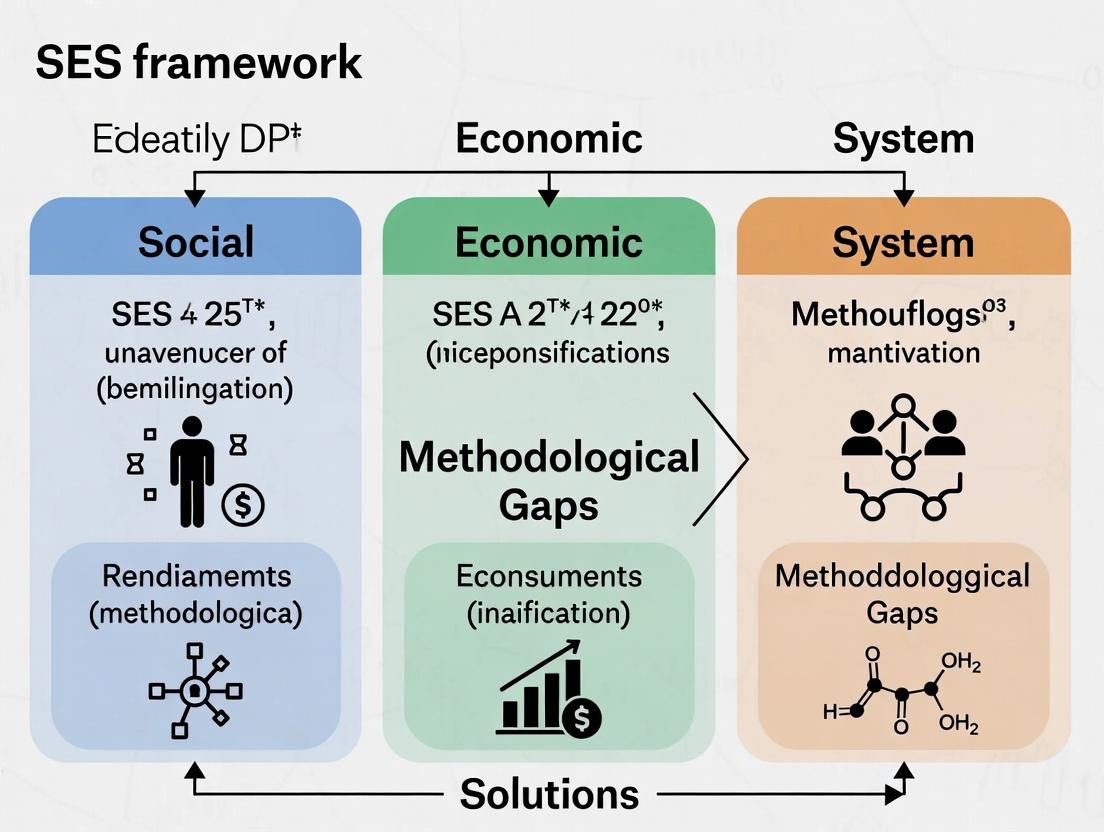
Understanding the SES Framework: Core Concepts, Evolution, and Current Relevance in Biomedical Research
Technical Support Center
Frequently Asked Questions (FAQs)
Q1: During in vitro SES (Safety, Efficacy, Specificity) profiling, my positive control for cytotoxicity consistently fails. What are the primary troubleshooting steps? A1: Follow this systematic protocol:
- Verify Reagent Integrity: Check the expiration date of your cytotoxic agent (e.g., Staurosporine). Reconstitute a fresh aliquot.
- Confirm Cell Health & Passage Number: Use low-passage cells (>90% viability via Trypan Blue exclusion). Over-passaged cells lose sensitivity.
- Optimize Concentration & Time: Re-establish the EC₉₀ kill curve. Use a 6-point, 1:3 serial dilution over 24-72 hours. See Table 1 for a standard reagent checklist.
- Validate Assay Readout: Ensure your detection reagent (e.g., ATP-based luminescence) is at room temperature, mixed thoroughly, and measured within the plate reader's linear range.
Q2: In target engagement assays, I observe high non-specific binding, skewing my specificity (S) metrics within the SES framework. How can I reduce this noise? A2: High background often stems from assay conditions.
- Optimize Blocking: Increase blocking time (overnight at 4°C) with a high-quality protein (e.g., 5% BSA in TBST) and include a mild detergent (e.g., 0.05% Tween-20).
- Implement Stringent Washes: Increase wash cycles (e.g., 6x post-primary antibody) and use a physiological buffer with correct ionic strength.
- Use Validated Controls: Include a competing ligand control (saturating unlabeled target ligand) to define specific signal. Include an isotype control for antibody-based detection. Data should be processed as: Specific Signal = Total Signal – Signal in Competing Ligand Well.
Q3: When integrating transcriptomic data for mechanistic efficacy (E) analysis, my pathway enrichment results are inconsistent. What methodological gaps should I address? A3: Inconsistency often relates to upstream bioinformatics.
- Normalization Method: Ensure raw RNA-seq counts are normalized using a robust method (e.g., DESeq2's median of ratios) appropriate for your study design. Do not rely solely on FPKM/TPM for differential expression.
- Gene Set Selection: Use curated, canonical pathways from sources like MSigDB's Hallmark sets. Avoid overly broad or redundant gene sets.
- Statistical Correction: Apply multiple testing correction (Benjamini-Hochberg FDR < 0.05) to enrichment p-values. Document your full computational protocol, including software versions.
Experimental Protocols
Protocol 1: Multi-Parametric High-Content Screening (HCS) for Concurrent SES Readouts
- Objective: Quantify efficacy (target translocation), specificity (off-target morphology), and safety (nuclear integrity) in a single live-cell experiment.
- Methodology:
- Seed U2OS cells expressing a fluorescently tagged target protein (e.g., GPCR-GFP) in a 96-well imaging plate.
- Treat with test compound (n=4), vehicle control, and reference controls (cytotoxic + target-active) for 24h.
- Stain with Hoechst 33342 (nuclei) and CellMask Deep Red (cytosol).
- Image using a high-content imager with 20x objective (4 sites/well).
- Analysis: Use HCS software to segment nuclei and cytoplasm. Calculate: Efficacy = GFP translocation coefficient (cytoplasmic/nuclear intensity ratio); Specificity = CellMask texture analysis (deviation from vehicle); Safety = Nuclear area and condensation index.
Protocol 2: Kinetic Target Engagement Assay (Cellular Thermal Shift Assay - CETSA)
- Objective: Provide direct, quantitative evidence of drug-target engagement (Efficacy pillar) in a cellular context.
- Methodology:
- Treat intact HEK293 cells (1e6 cells/mL) with compound or DMSO for 30 min.
- Aliquot cell suspensions into 8 PCR tubes (200 µL/tube).
- Heat each tube at distinct temperatures (e.g., 37°C to 67°C, 8-point gradient) for 3 min in a thermal cycler.
- Lyse cells by freeze-thaw, centrifuge (20,000 x g, 20 min).
- Analyze soluble fraction by Western blot or AlphaLISA for target protein.
- Analysis: Plot band intensity vs. temperature. Calculate the melting temperature (Tₘ) shift (ΔTₘ) between treated and vehicle samples. A positive ΔTₘ confirms target engagement.
Data Presentation
Table 1: Research Reagent Solutions for Core SES Assays
| Reagent Category | Specific Item (Example) | Function in SES Context | Critical Quality Check |
|---|---|---|---|
| Viability Probe | CellTiter-Glo 2.0 | Quantifies ATP as a marker of cell health (Safety pillar). | Confirm luminescent signal is linear from 100-10,000 cells/well. |
| Target Label | HaloTag-ligand TMR | Covalently labels HaloTag-fused target protein for localization & abundance studies (Efficacy/Specificity). | Validate labeling efficiency (>95%) via control cell line. |
| Pathway Reporter | pGL4.33[luc2P/SRE/Hygro] | Luciferase reporter for MAPK/ERK pathway activation (Efficacy pillar). | Test response to 10% FBS (positive control); Z'>0.5. |
| Positive Control (Cytotoxic) | Staurosporine | Induces apoptosis; serves as Safety assay control. | Confirm >80% cell death at 1 µM after 24h. |
| Positive Control (Target) | Known High-Affinity Ligand | Validates target engagement assay functionality (Efficacy pillar). | Its pIC₅₀ should be within 0.5 log of published value. |
Table 2: Example CETSA Data for Compound X
| Condition | Calculated Tₘ (°C) | ΔTₘ vs. Vehicle | p-value (t-test) | Interpretation |
|---|---|---|---|---|
| Vehicle (0.1% DMSO) | 52.1 ± 0.3 | -- | -- | Baseline target stability. |
| Compound X (1 µM) | 56.8 ± 0.5 | +4.7 °C | < 0.001 | Strong positive shift = high engagement. |
| Compound X (10 µM) | 58.2 ± 0.4 | +6.1 °C | < 0.001 | Dose-dependent stabilization. |
| Inactive Isomer (10 µM) | 52.4 ± 0.6 | +0.3 °C | 0.25 | No significant engagement. |
Mandatory Visualizations
SES Integrated Screening Workflow
Cellular Thermal Shift Assay (CETSA) Protocol
Key Pathways in PD-1/PD-L1 Drug Efficacy
Troubleshooting Guides & FAQs
Q1: Our in-vitro toxicity assay using 3D spheroids is showing high variability between batches. What are the key control points? A: Batch variability in 3D spheroids often stems from inconsistencies in cell aggregation and culture. Follow this protocol:
- Standardized Cell Preparation: Use a single-passage window (e.g., P4-P8). Count cells with an automated system after precise trypsinization quenching.
- Aggregation: Use ultra-low attachment, U-bottom 96-well plates. Centrifuge plates at 300 x g for 3 minutes immediately after seeding to initiate contact.
- Media & Supplement Control: Pre-warm all media to 37°C. Aliquot and freeze Matrigel or other ECM supplements in single-use volumes to avoid freeze-thaw cycles.
- Quantification Metric: Implement a high-content imaging system to measure spheroid diameter and circularity at 24h post-seeding. Discard batches where the coefficient of variation (CV) for diameter exceeds 15%.
Q2: When applying the SES (Safety, Efficacy, Sustainability) framework for early lead selection, how do we resolve conflicting data between predictive hepatotoxicity assays? A: Conflicting predictions between, for example, mitochondrial toxicity assays and phospholipidosis assays, indicate a gap in the integrated risk assessment. Implement a tiered experimental protocol:
- Tier 1 (Initial Screening): Run both assays in parallel using HepG2 cells.
- Tier 2 (Mechanistic Deconvolution): For compounds flagged in only one assay, proceed to primary human hepatocytes (PHHs) and measure more specific endpoints:
- For mitochondrial concern: Measure OCR (Oxygen Consumption Rate) and ECAR (Extracellular Acidification Rate) via Seahorse Analyzer.
- For phospholipidosis concern: Use high-content imaging with a lysosomal dye (e.g., LysoTracker) to quantify vesicle accumulation.
- Data Integration: Use the weighted scoring table below to make a go/no-go recommendation.
Q3: Our gene expression data for biomarker validation (e.g., KIM-1 for nephrotoxicity) is inconsistent across PCR platforms. How can we standardize this? A: Inconsistency typically arises from normalization and reagent issues.
- Normalization: Use a minimum of three validated reference genes (e.g., GAPDH, HPRT1, B2M) selected via geNorm or NormFinder algorithms. Do not rely on a single housekeeping gene.
- Reagent Calibration: Use a universal RNA calibrator sample across all runs and platforms. Create a standard curve for each assay to ensure PCR efficiency is between 90-110%.
- Protocol: Use 100ng total RNA input, reverse transcription with random hexamers and anchored oligo-dT primers, and pre-amplification if using low-abundance targets in a qPCR array.
Key Experimental Protocols
Protocol 1: Integrated Mitochondrial & Cytotoxicity Screening (Seahorse Assay) Objective: Simultaneously measure metabolic liability and cell death in real-time. Methodology:
- Seed HepaRG cells in Agilent Seahorse XF96 cell culture microplates at 40,000 cells/well.
- Incubate with test compound (5 concentrations, n=4) for 24h.
- One hour before assay, replace medium with XF Base Medium supplemented with 10mM glucose, 1mM pyruvate, and 2mM L-glutamine (pH 7.4). Incubate at 37°C, non-CO2.
- Load sensor cartridge with port injectors containing oligomycin (ATP synthase inhibitor, 1.5µM final), FCCP (uncoupler, 0.5µM final), and rotenone/antimycin A (Complex I/III inhibitors, 0.5µM final).
- Run the XF Cell Mito Stress Test assay on the Seahorse Analyzer.
- Immediately post-run, add CellTox Green dye to each well, incubate 15min, and measure fluorescence (485/520 nm) to quantify cytotoxicity.
Protocol 2: High-Content Imaging for Steatosis Assessment Objective: Quantify lipid accumulation in primary human hepatocytes. Methodology:
- Plate PHHs in 384-well collagen-coated imaging plates.
- Dose with compound for 72h with medium change at 48h.
- At endpoint, wash with PBS, fix with 4% paraformaldehyde for 20min.
- Wash, permeabilize with 0.1% Triton X-100 for 10min.
- Stain with LipidTOX Green (1:1000) and Hoechst 33342 (1µg/mL) for 1h.
- Image using a 20x objective on a high-content imager (e.g., ImageXpress Micro).
- Analyze: Identify nuclei via Hoechst channel, define cytoplasmic region, measure mean LipidTOX fluorescence intensity per cell. Report as fold-change over vehicle control.
Data Tables
Table 1: Comparative Performance of Predictive Hepatotoxicity Assays
| Assay Platform | Target Pathway | Key Metric (Typical Threshold) | Concordance with Clinical DILI (Literature %) | Throughput | Cost per Compound |
|---|---|---|---|---|---|
| 2D HepG2 Cytotoxicity | General cytotoxicity | IC50 (<100 µM) | ~50-60% | High | Low |
| 3D Spheroid (HepG2/HepaRG) | Metabolic function, chronic toxicity | Viability (Selectivity Index <10) | ~70-75% | Medium | Medium |
| Mitochondrial Toxicity (Seahorse) | Oxidative phosphorylation | Basal OCR inhibition (>25%) | ~70% | Low-Medium | High |
| Transporter Inhibition (CYP450, BSEP) | Drug metabolism & efflux | IC50 (<10 µM) | High for specific DILI | High | Medium |
| High-Content Imaging (PHHs) | Multiple (steatosis, stress) | Multiplexed readouts (Z-score >2) | ~75-80% | Low | High |
Table 2: Weighted Scoring for SES Lead Selection in Conflicting Toxicity Data
| Assay Category | Assay Result | Assay Concordance Weight (1-3) | Clinical Severity Weight (1-3) | Composite Score (Result x Concordance x Severity) |
|---|---|---|---|---|
| Mitochondrial Dysfunction (Seahorse) | Positive (1) or Negative (0) | 3 (High translational concordance) | 3 (High risk of serious DILI) | Score = Result * 9 |
| Phospholipidosis (HCS) | Positive (1) or Negative (0) | 2 (Moderate concordance) | 2 (Moderate risk, often reversible) | Score = Result * 4 |
| Genomic Biomarker (e.g., KIM-1) | Upregulated (1) or Not (0) | 2 (Emerging biomarker) | 3 (High specificity) | Score = Result * 6 |
| Total Lead Risk Score | Sum of all Composite Scores | Go Decision: Total Score < 5 |
Visualizations
SES Framework Lead Selection Workflow
Key Mitochondrial Toxicity Signaling Pathways
The Scientist's Toolkit: Research Reagent Solutions
| Reagent/Material | Function & Rationale |
|---|---|
| Primary Human Hepatocytes (PHHs) | Gold standard for hepatotoxicity assessment due to full complement of human drug-metabolizing enzymes and transporters. Cryopreserved, plateable formats enable reproducible use. |
| HepaRG Cell Line | Bipotent progenitor cell that differentiates into hepatocyte-like and biliary-like cells. Maintains expression of key CYPs (e.g., CYP3A4) and nuclear receptors, offering a balance of relevance and throughput. |
| Matrigel / BME (Basement Membrane Extract) | Used for 3D culture and sandwich cultures of hepatocytes. Maintains polarized morphology, enhances longevity, and supports albumin/Urea synthesis. |
| Seahorse XFp/XFe96 Analyzer Kits | Pre-optimized kits (Mito Stress Test, Glycolysis Test) for real-time, label-free measurement of metabolic function in live cells, critical for mitochondrial liability screening. |
| LipidTOX (HCS Lipid Stain) | Neutral lipid stain optimized for high-content screening. Provides high specificity and signal-to-noise ratio for quantifying steatosis (fatty liver) in fixed cells. |
| LC-MS/MS Grade Solvents & Stable Isotope Standards | Essential for generating high-quality metabolomics and proteomics data to identify novel toxicity biomarkers within the SES framework. |
| Multi-Plex Cytokine/KIM-1 Assay (MSD or Luminex) | Enables quantitative measurement of multiple injury biomarkers (e.g., KIM-1, IL-6, IL-8) from a single small-volume supernatant sample, enhancing translational safety assessment. |
Technical Support Center: Troubleshooting SES Framework Implementation
Support Context: This center provides guidance for researchers implementing Systematic, Empirical, and Scientific (SES) principles in biomedical research, specifically within drug development. It addresses common methodological gaps identified in ongoing thesis research on SES framework robustness.
Troubleshooting Guides
Issue 1: Non-Systematic Experimental Design Leading to Irreproducible Results
- Problem: High variability in assay outputs between operators or lab sites.
- Diagnosis: Lack of a Standard Operating Procedure (SOP) with defined tolerances for key variables.
- Solution: Implement a pre-experimental checklist.
- Protocol: 1. Document all reagent lot numbers and calibration dates for instruments. 2. Pre-define acceptance criteria for control sample values. 3. Use a randomized sample processing order to minimize batch effects. 4. Require a pilot run with n=3 for any new assay configuration before full experiment.
Issue 2: Empirical Data Collection Insufficient for Robust Statistical Analysis
- Problem: Inability to achieve statistical significance (p < 0.05) despite observed phenotypic effects.
- Diagnosis: Underpowered experiments due to low sample size (n).
- Solution: Conduct an a priori power analysis.
- Protocol: 1. From pilot data or literature, estimate the expected effect size (e.g., Cohen's d) and data variance. 2. Set desired statistical power (typically 0.8 or 80%). 3. Use software (e.g., G*Power) to calculate the minimum required sample size. 4. Adjust experimental design to meet this n, potentially using higher-throughput methods.
Issue 3: Failure to Adhere to Scientific Principles of Falsifiability
- Problem: Inability to distinguish true target engagement from off-target effects in cell-based assays.
- Diagnosis: Lack of appropriate controls to challenge the hypothesis.
- Solution: Integrate orthogonal and negative controls.
- Protocol: 1. Orthogonal Control: Confirm a key finding using a different methodological principle (e.g., confirm Western blot protein level change with targeted mass spectrometry). 2. Negative Control: Use a genetically modified (CRISPR knockout) or pharmacologically inhibited (specific inhibitor) system to demonstrate the effect is abolished when the hypothesized target is absent/inactive.
Frequently Asked Questions (FAQs)
Q1: What is the minimum dataset required to claim an observation is "empirical" within the SES framework? A: An empirical claim must be supported by quantitative data from at least three independent experimental replicates (biological, not just technical), collected under systematically controlled conditions. The dataset must allow for the calculation of a measure of central tendency (mean/median) and variance (SD/SEM). See Table 1.
Q2: How do I systematically document an unexpected finding (serendipity) without compromising the integrity of my planned experiment? A: Follow the "Observe, Document, Hypothesize, Test" protocol. 1. Observe & Document: Immediately note the anomaly with timestamp, conditions, and raw image/data. Do not alter the primary experiment. 2. Hypothesize: Formulate a testable hypothesis for the cause after the planned experiment concludes. 3. Test: Design a new, systematic experiment explicitly to test this new hypothesis, including proper controls.
Q3: My assay is inherently variable (e.g., primary cell assays). How can I apply systematic principles? A: Systematism controls for what can be controlled and characterizes what cannot. Implement: 1) Standardized donor/source criteria, 2) Rigorous passage number limits, 3) Internal reference controls in every run (e.g., a standard agonist response), and 4) Clear acceptance criteria for control performance. The empirical data must then include this characterized variability in its error bars and statistical models.
Table 1: Empirical Data Thresholds for Common Assay Types
| Assay Type | Minimum Biological Replicates (n) | Recommended Statistical Test | Primary Data to Report |
|---|---|---|---|
| Cell Viability (MTT/CTG) | 4 (per condition) | Two-way ANOVA with post-hoc | Mean % viability ± SEM, raw absorbance/luminescence values |
| qPCR (Gene Expression) | 3 independent samples | ΔΔCt method, Student's t-test | Ct values, reference genes used, fold-change ± SD |
| Western Blot Densitometry | 3 independent blots | Non-parametric Mann-Whitney U test | Representative blot, normalized band intensity ± SEM |
| In Vivo Efficacy Study | 6-8 animals per group | Mixed-effects model | Individual animal data points, mean tumor volume/score ± SEM |
Table 2: Common Methodological Gaps & SES-Compliant Solutions
| Identified Gap | Systematic Principle Solution | Empirical Validation Needed |
|---|---|---|
| Unblinded analysis | Implement sample coding; automated data processing scripts. | Compare outcomes from blinded vs. unblinded analysis on a pilot set (n=5). |
| Subjective endpoint scoring | Use pre-defined, quantitative scoring rubric; employ two independent scorers. | Calculate Inter-rater reliability (Cohen's Kappa) report Kappa > 0.7. |
| Uncontrolled environmental factors | Log ambient CO2, temperature, humidity in lab; use equipment timers. | Correlate control sample performance with logged factors over 30 runs. |
Experimental Protocol: SES-Compliant Dose-Response Analysis
Objective: To determine the IC50 of a novel compound on cancer cell proliferation systematically, empirically, and scientifically.
Methodology:
- Systematic Setup:
- Seed cells in 96-well plates using an automated dispenser. Include background (media only), vehicle control (DMSO), and positive control (reference inhibitor) wells on every plate.
- Prepare a 10-point, half-log dilution series of the test compound in triplicate.
- Randomize the plate layout using online software to avoid positional effects.
Empirical Measurement:
- After 72h incubation, add CellTiter-Glo reagent per manufacturer's instructions.
- Measure luminescence on a plate reader. Export raw data (RLU values).
Scientific Analysis:
- Normalize data: (Sample - Background Median) / (Vehicle Control Median - Background Median) * 100%.
- Fit normalized data to a sigmoidal dose-response model (e.g., 4-parameter logistic) using software (e.g., GraphPad Prism).
- Report the calculated IC50 value with 95% confidence interval, the Hill slope, and the R² of the fit. The model must be falsifiable—if R² < 0.9, the fit is rejected.
Pathway & Workflow Diagrams
SES Research Iterative Workflow
RTK-PI3K-AKT-mTOR Pathway & Inhibition
The Scientist's Toolkit: Research Reagent Solutions
| Reagent / Material | Function in SES Context | Key Consideration for Systematism |
|---|---|---|
| CRISPR/Cas9 Knockout Cell Lines | Provides isogenic negative controls to test target specificity (Scientific Principle of Falsifiability). | Validate knockout via sequencing (DNA), Western blot (protein), and functional assay. Use early passage aliquots. |
| Cryopreserved Primary Cells (e.g., HUVEC, HPMC) | Provides biologically relevant empirical data beyond immortalized cell lines. | Document donor characteristics and passage number. Always include a viability assay post-thaw; set a minimum acceptance threshold (e.g., >85%). |
| Validated Chemical Probes (e.g., from SGC) | High-quality tool compounds with published data on selectivity and use. Enables systematic comparison. | Source from reputable suppliers. Use at recommended concentrations. Include matched inactive analogs as controls if available. |
| Internal Control Reference Standards (e.g., assay-ready plates) | Allows for inter-experiment and inter-operator normalization, enabling systematic data aggregation. | Run in every experimental batch. Track performance over time via a control chart to detect assay drift. |
| Sample Anonymization / Blinding Software | Removes subjective bias during data collection and analysis, upholding scientific objectivity. | Implement before data generation. Document the blinding key separately. Unblind only after final analysis is locked. |
The Central Role of SES in Modern Regulatory Submissions (e.g., FDA, EMA)
Technical Support Center
Troubleshooting Guide & FAQs
Q1: Our Study Data Tabulation Model (SDTM) datasets are being rejected by the FDA's Technical Rejection Criteria. The error cites invalid SES (Subject Elements) domains. What is the most common cause? A1: The most common cause is a mismatch between the SESTDTC (Subject Element Start Date/Time) and the study reference period or the RFSTDTC (Subject Reference Start Date/Time) in the Demographics (DM) domain. Ensure every subject's first SES start date (e.g., first treatment, first exposure) is logically aligned with their reference start date. The SES domain must precisely anchor subject milestones to the study timeline.
Q2: How should we handle protocol deviations that alter subject status (e.g., a temporary halt in dosing) within the SES framework for an EMA submission? A2: Create a new SES record for the new status. Do not modify the original SES record. For a dosing halt:
- Record 1:
SESTESTCD = TREATMENT,SESCAT = ASSIGNED,SESPRESP = Y,SESOCCUR = Y. - Record 2 (upon halt):
SESTESTCD = TREATMENT,SESCAT = INTERRUPTED,SESPREsp = N,SESOCCUR = Y. - Record 3 (upon resumption): A new record with
SESCAT = RE-ASSIGNED. This creates an audit trail of subject element states critical for review.
Q3: Our electronic submission (eCTD) to the FDA passed validation but received a major comment that the "analysis population derivation is not traceable from SES and SE (Subject Elements)." How do we fix this?
A3: This indicates a gap between the SES-defined states and the Analysis Data Model (ADaM) population flags (e.g., SAFFL, ITTFL, EFFFL). The solution is to provide a clear derivation protocol (see below) and ensure every population flag in ADSL can be directly linked to a rule based on SES/SE variables (SESCAT, SESOCCUR, SEENDTC).
Q4: For a complex oncology trial with multiple treatment cycles and dose modifications, how granular should SES records be?
A4: Extremely granular. Each distinct treatment element (e.g., Induction Cycle 1, Maintenance Cycle 2 at Reduced Dose) must be a separate record. Use SESCAT (e.g., "INDUCTION", "MAINTENANCE"), SESSCAT (e.g., "CYCLE1", "CYCLE2"), and SESSPID to uniquely identify and order elements. This granularity is essential for accurate time-to-event analyses and safety reviews.
Detailed Methodology: Linking SES to Analysis Populations
Protocol Title: Derivation of ADaM Analysis Population Flags from Subject Elements Data.
Objective: To provide a reproducible, traceable methodology for deriving regulatory analysis populations (Safety, Intent-to-Treat, Efficacy) based on subject participation states defined in the SES domain.
Materials & Software:
- Source SDTM datasets: DM, SE, SES, EX (Exposure).
- ADaM dataset specification: ADSL (Subject-Level Analysis Dataset).
- Statistical computing environment (e.g., SAS, R).
- CDISC conformance rules v1.7 or later.
Procedure:
- Identify Key Subject Elements: Collaborate with the study team to define the critical elements that determine trial participation (e.g., "Randomized," "Treated," "Completed Week 4 Visit," "Major Protocol Deviation X").
- Map to SES: Ensure each key element is represented as a record in the SES domain with appropriate
SESTESTCD,SESTEST,SESCAT, and a clearSESPRESP(Planned) andSESOCCUR(Actual) value. - Derivation Logic for ADSL Flags:
- Safety Population (SAFFL):
SAFFL = 'Y'if(SESTESTCD = 'TREATMENT' and SESCAT = 'ASSIGNED' and SESOCCUR = 'Y'). This confirms the subject received at least one dose of study treatment. - Intent-to-Treat Population (ITTFL):
ITTFL = 'Y'if(SESTESTCD = 'RANDOMIZATION' and SESOCCUR = 'Y'). This confirms the subject was randomized. - Efficacy Population (EFFFL):
EFFFL = 'Y'ifITTFL = 'Y'AND(SESTESTCD = 'WEEK4VISIT' and SESOCCUR = 'Y')AND(SESTESTCD = 'MAJORPROTDEV' and SESOCCUR = 'N'). This is study-specific, often requiring completion of a key milestone without a critical deviation.
- Safety Population (SAFFL):
- Documentation: Create a Define.xml appendix or an Analysis Results Metadata (ARM) document that explicitly links each ADSL population flag derivation to the specific SES records and logic used. Include this as part of the submission package.
Validation: Perform QC checks by sampling subjects and manually verifying that their SES records lead to the correct population flag assignments in ADSL.
Table 1: Common SES-Related Issues in FDA Submissions (2022-2023)
| Issue Category | Frequency (%) | Typical Resolution Time (Weeks) |
|---|---|---|
| SES/SE Timing Inconsistencies | 42% | 2-4 |
| Incomplete Subject Element States | 28% | 4-6 |
| Poor Traceability to Analysis Populations | 18% | 6-8 |
| Invalid SESCAT/SESSCAT Codelist Usage | 12% | 1-2 |
Table 2: Impact of Robust SES Implementation on Submission Quality
| Metric | Submissions with Minimal SES Gaps | Submissions with Major SES Gaps |
|---|---|---|
| First-Pass Acceptance Rate* | 85% | 35% |
| Average Review Cycle Questions | 12 | 47 |
| Time to Approval (Months) | 10.2 | 16.8 |
*Acceptance without a Refuse-to-File or Major Amendment request.
Visualizations
SES Drives End-to-End Submission Integrity
Population Flag Derivation Logic Flow
The Scientist's Toolkit: Research Reagent Solutions for SES Implementation
Table 3: Essential Tools for Robust SES Framework Implementation
| Item | Function/Benefit | Example/Note |
|---|---|---|
| CDISC SDTM/ADaM IG | The foundational rulebook. Provides standard variables, structures, and examples for implementing SES/SE domains. | CDISC Published Guides v3.4 & v1.3. |
| Controlled Terminology (CT) | Pre-defined codelists for SESTESTCD, SESCAT, etc. Ensures consistency and regulatory acceptance. |
NCI EVS CT, including latest "SUBJECT ELEMENTS" terms. |
| Metadata Repository | A centralized system (e.g., using Define.xml) to document the origin and purpose of each SES record. Enforces traceability. | OpenStudyBuilder, PHUSE CSR Template. |
| Automated Consistency Checks | Scripts (SAS/R/Python) to validate SES timing against DM/EX and flag logical gaps pre-submission. | Custom programs checking SESTDTC >= RFSTDTC. |
| Therapeutic Area (TA) User Guide | TA-specific guidance (e.g., CDISC Oncology, Vaccines) on common subject elements and their representation in SES. | Informs granular SESCAT values (e.g., "RUN-IN", "CROSSOVER"). |
Technical Support Center: Troubleshooting SES Framework Research
Frequently Asked Questions (FAQs)
Q1: Our preclinical study in a genetically uniform mouse model showed high efficacy, but the drug failed in Phase II human trials with a more socioeconomically diverse population. What could be the primary SES-related methodological gap?
A1: This failure likely stems from the SES-Exposure Gap. Preclinical models (e.g., inbred mice in controlled environments) lack the variable environmental exposures (diet, pollutants, stress) correlated with SES in humans. These exposures can drastically alter drug metabolism pathways (e.g., CYP450 enzyme activity) and disease pathophysiology. Your model failed to account for this biological embedding of SES.
Q2: We are designing a biomarker validation study. How can we avoid "SES Biomarker Confounding" where our putative biomarker is actually a proxy for nutritional status or access to care?
A2: Implement Multivariate Stratified Sampling. Actively recruit and stratify participants not just by disease stage, but by key SES dimensions (income, education, ZIP code-derived ADI). During analysis, use multiple regression to statistically control for these factors and confirm the biomarker's independent predictive value. See Protocol 1 below.
Q3: Our cell culture work uses standard fetal bovine serum (FBS). Could this introduce an SES analog bias in translational research?
A3: Yes. Standard FBS represents a single, uniform, and affluent "nutritional environment" not reflective of human variation. Cells cultured this way may develop metabolic dependencies not present in cells from organisms under nutritional stress. Consider experiments supplementing media with variable nutrient cocktails to mimic metabolic states found across SES gradients.
Q4: In retrospective data analysis, how do we handle missing SES data in electronic health records (EHR), which is often non-random?
A4: Do not simply exclude cases with missing SES data. Employ Multiple Imputation with Sensitivity Analysis. Use known variables (insurance type, neighborhood data, diagnosis codes) to impute missing SES values. Run your analysis on multiple imputed datasets and perform a sensitivity analysis to see if conclusions hold under different assumptions about the missing data mechanism.
Troubleshooting Guides
Issue: Inconsistent Drug Response in Population-Based Cohort
- Symptoms: High inter-individual variability, subgroup analyses show correlation with zip code or education level.
- Diagnosis: SES-Driven Biological Heterogeneity未被控制。
- Solution: Apply Covariate Adjustment with Propensity Scoring. 1) Collect granular SES data (see Toolkit). 2) Generate a propensity score for being in a low-SES group based on covariates. 3) Match participants or use the score as a covariate in dose-response modeling. This isolates the drug's effect from SES-confounders.
- Validation Experiment: Re-analyze response data with and without SES adjustment. A stable effect size after adjustment indicates robust findings.
Issue: Animal Model Fails to Replicate Human Disease Progression Pattern
- Symptoms: Pathological markers appear in a different sequence or magnitude than in human patient biopsies.
- Diagnosis: Overly Simplistic Model Environment ignoring the multifactorial stress exposures of low-SES.
- Solution: Implement a Chronic Variable Stress (CVS) Paradigm in rodents alongside your disease model. See Protocol 2.
- Validation: Compare transcriptomic profiles from your CVS-model animals to human data from low-SES patient biopsies (from public repositories). Overlap in stress and disease pathways validates the model's translational relevance.
Experimental Protocols
Protocol 1: Controlling for SES in Biomarker Validation Studies
Objective: To isolate the predictive value of a novel inflammatory biomarker (e.g., Novel Inflammatin X) for cardiovascular event risk, independent of SES.
Cohort Recruitment (N=1000): Recruit participants with elevated baseline risk. Actively stratify recruitment to ensure balanced representation across four SES quadrants defined by:
- High Education/High Income
- High Education/Low Income
- Low Education/High Income
- Low Education/Low Income
- Use census tract data (Area Deprivation Index) as a secondary, objective measure.
Data Collection:
- Biomarker: Plasma sample analyzed via ELISA for Novel Inflammatin X.
- Clinical Endpoint: Time-to-first major adverse cardiovascular event (MACE) over 5 years.
- Covariates: Age, sex, BMI, smoking status, insurance type, self-reported stress (PSS scale), dietary quality index.
Statistical Analysis:
- Perform Cox Proportional Hazards regression.
- Model 1: Biomarker level only.
- Model 2: Model 1 + traditional clinical covariates.
- Model 3: Model 2 + SES covariates (education, income, ADI, insurance).
- Interpretation: If the hazard ratio for the biomarker remains significant and stable from Model 2 to Model 3, it is robust to SES confounding.
Protocol 2: Incorporating SES-Relevant Stress in a Rodent Metabolic Disease Model
Objective: To induce metabolic heterogeneity in C57BL/6 mice mimicking SES-linked health disparities, for testing a diabetic therapeutic.
- Animals: 8-week-old male C57BL/6 mice (n=40).
- Groups (n=10/group):
- Control: Standard chow, normal housing.
- Diet-Induced Obesity (DIO): High-fat diet (60% kcal from fat).
- Chronic Variable Stress (CVS): Standard chow, subjected to a different mild stressor daily (e.g., damp bedding, cage tilt, white noise, temporary social isolation) on an unpredictable schedule for 8 weeks.
- DIO+CVS: High-fat diet + CVS protocol.
- Weekly Monitoring: Body weight, fasting blood glucose.
- Terminal Analysis (Week 16): Oral glucose tolerance test (OGTT), plasma insulin, corticosterone, hepatic lipid content, hypothalamic RNA-seq for stress and metabolic pathways.
- Therapeutic Arm: After 8 weeks of phenotype establishment, subdivide each group (n=5) into vehicle vs. drug treatment for efficacy testing.
Data Presentation
Table 1: Impact of SES Covariate Adjustment on Biomarker Hazard Ratios (Simulated Data)
| Biomarker | Model 1 (Unadjusted) HR [95% CI] | Model 2 (Clinical Covariates) HR [95% CI] | Model 3 (+SES Covariates) HR [95% CI] | Conclusion |
|---|---|---|---|---|
| Novel Inflammatin X | 2.5 [1.8-3.4] | 2.3 [1.6-3.2] | 2.2 [1.5-3.1] | Robust. Slight attenuation, remains significant. |
| Plasma Vitamin D | 0.6 [0.5-0.8] | 0.7 [0.5-0.9] | 0.9 [0.7-1.2] | Confounded. Effect nullified after SES adjustment. |
| CRP (Standard) | 1.8 [1.3-2.5] | 1.6 [1.1-2.2] | 1.5 [1.0-2.1] | Partially Confounded. Confidence interval widens to include 1.0. |
Table 2: Metabolic Phenotypes in SES-Mimetic Mouse Model (Example Outcomes)
| Group | Final Body Weight (g) | OGTT AUC (mmol/L*min) | Fasting Corticosterone (ng/mL) | Hepatic Steatosis Score |
|---|---|---|---|---|
| Control | 28.5 ± 1.2 | 1200 ± 150 | 50 ± 15 | 1.0 ± 0.3 |
| DIO Only | 45.2 ± 3.1* | 2800 ± 300* | 65 ± 20 | 3.5 ± 0.5* |
| CVS Only | 30.1 ± 1.5 | 1500 ± 200# | 180 ± 30*# | 1.8 ± 0.4# |
| DIO + CVS | 48.8 ± 2.8* | 3500 ± 400*# | 220 ± 40*# | 4.5 ± 0.6*# |
*p<0.05 vs Control, #p<0.05 vs DIO Only. Data presented as mean ± SD.
Visualizations
Diagram 1: SES Gaps Impact on Research Translation Pathway
Diagram 2: Chronic Variable Stress (CVS) Experimental Workflow
The Scientist's Toolkit: Research Reagent Solutions for SES-Aware Research
| Item | Function in SES Context | Example/Supplier |
|---|---|---|
| Area Deprivation Index (ADI) Data | Objective Neighborhood SES Metric. Geocodes participant addresses to a percentile-ranked index of socioeconomic disadvantage. Controls for environmental confounders. | University of Wisconsin School of Medicine Public Health. |
| Variable Nutrient Media | Models Nutritional Inequality. Base media supplemented with different fatty acid ratios, micronutrient levels, or "serum" from donors of varying health status to mimic diverse human diets. | Custom formulation from providers like Sigma; HyClone Characterized FBS variants. |
| Chronic Variable Stress (CVS) Protocol Kit | Standardizes stress induction in rodents to simulate the psychosocial stress burden associated with low SES. Increases translational face validity. | Detailed protocols from SCOPUS/PubMed; stressors from lab supply companies. |
| Multiplex ELISA for Stress & Inflammation | Measures intertwined pathways. Panels quantifying cortisol/corticosterone, CRP, IL-6, TNF-α, and metabolic hormones (insulin, leptin) from a single sample to capture SES-linked biology. | Meso Scale Discovery, Luminex, Abcam. |
| Data Imputation Software (e.g., R 'mice') | Handles missing SES data. Uses multiple imputation to address non-random missingness in EHR-derived SES variables, reducing selection bias. | R package 'mice'; STATA ICE. |
| Propensity Score Matching Packages | Balances comparison groups. Statistically creates matched cohorts that are equivalent on observed SES covariates, isolating the variable of interest. | R 'MatchIt'; Python 'PropensityScoreMatching'. |
Identifying Critical SES Framework Gaps: Data, Analysis, and Real-World Application Challenges
Troubleshooting Guides & FAQs
Q1: Our multi-omics data integration failed due to mismatched gene identifiers from different sources. What is the first step to resolve this? A: The primary issue is inconsistent naming conventions. Implement a robust identifier mapping pipeline. First, audit all data sources for their native ID types (e.g., Ensembl ID, Entrez ID, gene symbol). Use a centralized, version-controlled mapping service like the HGNC (HUGO Gene Nomenclature Committee) or UniProt for proteins. Convert all identifiers to a single, stable standard (e.g., Ensembl Gene ID v110) before integration. Common pitfalls include assuming gene symbol uniqueness and not accounting for identifier version deprecation.
Q2: When annotating clinical phenotypes, our team uses different terms for the same condition (e.g., "Stage III" vs. "Advanced"). How can we enforce consistency? A: Adopt a formal clinical ontology. For oncology, implement the NCI Thesaurus (NCIt) or SNOMED CT. Establish a pre-experiment protocol where all clinical data annotators must select terms from a pre-defined, project-specific subset (a "slim") of the chosen ontology. Use ontology management software (e.g., Protégé) to create and enforce this controlled vocabulary. Inconsistencies post-collection require manual reconciliation against the ontology, which is time-consuming.
Q3: Cell line contamination or misidentification is skewing our meta-analysis. How can we prevent this? A: This is a critical data provenance gap. Mandate the following steps: 1) Authentication: Use STR profiling for all human cell lines at the start and end of experiments. 2) Standardized Nomenclature: Report cell lines using the Cellosaurus accession ID (e.g., CVCL_0030 for A549). 3) Metadata Reporting: In your methods, always detail the source repository (e.g., ATCC), passage number, and mycoplasma testing status. Never use colloquial or lab-specific names in published data.
Q4: Pathway analysis results are irreproducible between tools (e.g., DAVID vs. Reactome). What parameters should we standardize? A: The discrepancy often stems from different underlying pathway databases and statistical models. Standardize your workflow: 1) Gene Set Source: Commit to one database (e.g., Reactome, GO, KEGG) and note its version. 2) Background List: Use the same genomic background (e.g., all protein-coding genes from ENSEMBL) for all analyses. 3) Correction Method: Consistently apply a multiple testing correction (e.g., Benjamini-Hochberg FDR < 0.05). Documenting these three parameters is essential for reproducibility.
Q5: How do we handle legacy data from older studies that lack any ontological annotation? A: Create a retrospective curation pipeline. This involves: 1) Data Audit: Inventory all data fields and free-text entries. 2) Term Mapping: Use text-mining tools (e.g., OBO Annotator, MetaMap) to suggest mappings to current ontologies like EFO (Experimental Factor Ontology). 3) Expert Review: Have a domain expert validate all automated mappings. 4) Flagging: Clearly mark retrospectively curated data in your metadata with a "curationdate" and "curationmethod" field. Do not alter the original raw data file.
Table 1: Impact of Inconsistent Standardization on Meta-Analysis Reproducibility
| Study Feature Lacking Standardization | % of Studies Affected (2020-2024 Sample)* | Average Delay in Data Reuse (Weeks) | Risk of False Positive/False Negative Conclusion |
|---|---|---|---|
| Cell Line Identification (no STR/CRISPR) | 23% | 3-4 | High |
| Gene/Protein Identifier (mixed sources) | 65% | 2-3 | High |
| Clinical Phenotype (free-text only) | 41% | 4-6 | Medium-High |
| Experimental Protocol (incomplete MIAME/ARRIVE) | 58% | 2-5 | Medium |
| Units of Measurement (unclear or missing) | 19% | 1-2 | Medium |
Data synthesized from recent reviews in *Nature Scientific Data and Bioinformatics.
Table 2: Adoption Rates of Key Ontologies in Public Repositories (2023)
| Ontology | Domain | Use in ArrayExpress (%) | Use in GEO (%) | Mandated by Major Journal? |
|---|---|---|---|---|
| Cell Ontology (CL) | Cell Types | 78% | 62% | Partial |
| Experimental Factor Ontology (EFO) | Experimental Variables | 85% | 70% | Yes (EMBL-EBI) |
| Disease Ontology (DOID) | Human Diseases | 71% | 58% | Partial |
| Gene Ontology (GO) | Gene Function | 95% | 92% | Yes (widespread) |
| Units of Measurement Ontology (UO) | Quantities | 45% | 32% | No |
Detailed Experimental Protocols
Protocol 1: Implementing a Unified Data Processing Pipeline for Transcriptomics Meta-Analysis
Objective: To harmonize raw RNA-Seq data from disparate studies for integrated differential expression analysis.
Materials: High-performance computing cluster, Docker/Singularity, FastQC (v0.12.1), MultiQC (v1.14), nf-core/rnaseq pipeline (v3.12), Ensembl reference genome & annotation (v110), sample metadata sheet (.tsv).
Method:
- Data Audit & Metadata Curation: Collect all study SRA Run IDs. Manually curate a metadata table with standardized fields:
sample_id,study_id,condition(EFO term),organism(NCBI TaxID),sex,cell_type(CL term),instrument. Validate with ISAcreator tools. - Containerized Workflow: Use the nf-core/rnaseq pipeline via Docker to ensure identical software versions. Command:
nextflow run nf-core/rnaseq --input samplesheet.csv --genome GRCh38 --outdir results. - Standardized Reference: Align all samples to the same version of the human reference genome (GRCh38) using STAR. Quantify reads against the same gene annotation (Ensembl v110) using Salmon.
- Identifier Harmonization: The pipeline outputs gene-level counts using stable Ensembl Gene IDs. This serves as the common denominator.
- Batch Effect Assessment: Use the
limma::removeBatchEffect()function in R to visualize and correct for technical variation between studies before downstream analysis.
Protocol 2: Retrospective Ontological Annotation of Clinical Trial Datasets
Objective: To map free-text clinical observations from historical studies to standardized ontology terms.
Materials: Dataset in CSV format, OLS (Ontology Lookup Service) API, Zooma annotation tool (EMBL-EBI), R or Python environment with ontologyIndex and jsonlite packages.
Method:
- Vocabulary Extraction: Isolate all unique strings from relevant clinical columns (e.g.,
diagnosis,response,adverse_event). - Automated Mapping: Submit the unique string list to the Zooma service via its REST API. Zooma will return suggested mappings to terms from ontologies like SNOMED CT, NCIt, and EFO, with a confidence score.
- Curation & Validation: Export results to a spreadsheet. A clinical expert must review each mapping, especially those with confidence < 95%. Accept, reject, or provide a manual mapping.
- Metadata Enhancement: Create a new column in the dataset (e.g.,
diagnosis_ontology_id) populated with the curated ontology term ID (e.g.,NCIT:C3493for 'Stage III Colon Cancer'). - Provenance Logging: Create an accompanying log file documenting the curation date, curator name, Zooma version, and rules for manual overrides.
Diagrams
Title: SES Framework Data Standardization Workflow
Title: Ontology Mapping & Curation Process
The Scientist's Toolkit: Research Reagent Solutions
Table 3: Essential Resources for Data Standardization
| Item | Function in Standardization | Example/Supplier |
|---|---|---|
| Cellosaurus | Provides unique, stable accession IDs (CVCL_) for cell lines, crucial for unambiguous reporting and preventing misidentification. | https://web.expasy.org/cellosaurus/ |
| Ensembl Gene ID | A stable, versioned identifier system for genes across species. Serves as a reliable key for cross-dataset integration. | ENSG00000139618 (Human BRCA2) |
| Experimental Factor Ontology (EFO) | A structured ontology for describing experimental variables, treatments, and phenotypes in bioscience. Critical for metadata annotation. | https://www.ebi.ac.uk/efo/ |
| ISA-Tab Format & Tools | A general-purpose framework for representing experimental metadata using investigation, study, and assay files. Ensures complete metadata capture. | ISAcreator software suite |
| BioContainers | Provides versioned, containerized bioinformatics tools (Docker/Singularity). Eliminates "works on my machine" issues and ensures pipeline reproducibility. | https://biocontainers.pro/ |
| Ontology Lookup Service (OLS) | A centralized repository and API for querying hundreds of biomedical ontologies. Enables real-time term lookup and validation. | https://www.ebi.ac.uk/ols4 |
Technical Support Center
Frequently Asked Questions (FAQs)
Q1: In our observational study of socioeconomic status (SES) and a specific health outcome, how can we determine if the relationship is causal or confounded? A1: Establishing causality requires rigorous design. First, map potential confounders (e.g., age, ethnicity, access to care, environmental factors) using a DAG. For analysis, consider propensity score matching (PSM) to create balanced groups or use instrumental variable (IV) analysis if a suitable variable (e.g., policy changes, genetic instruments) is available. Always report the assumptions and limitations of your chosen method.
Q2: Our matched cohort study shows residual bias after PSM. What are the primary troubleshooting steps? A2: Residual bias often indicates poor overlap or unmeasured confounding.
- Check Overlap: Generate a love plot of standardized mean differences (SMDs) for all covariates pre- and post-matching. Target SMD < 0.1.
- Assess Balance: Conduct hypothesis tests (e.g., t-tests) on covariates post-matching; they should be non-significant.
- Sensitivity Analysis: Perform a Rosenbaum bounds analysis to quantify how strong an unmeasured confounder would need to be to alter your inference.
Q3: We are using an instrumental variable (IV) to estimate causal effect, but the F-statistic from the first-stage regression is low (F=3.5). What does this mean and how do we proceed? A3: A low F-statistic (<10) indicates a "weak instrument," which can cause severe bias. You must:
- Find a Stronger Instrument: Re-evaluate your IV's theoretical and empirical relevance.
- Report Diagnostics: Always report the first-stage F-statistic and partial R².
- Use Robust Methods: Switch to limited information maximum likelihood (LIML) or Fuller estimation, which are less biased with weak instruments than two-stage least squares (2SLS).
- Acknowledge Limitation: Clearly state that causal estimates may be unreliable due to instrument weakness.
Q4: How do we handle time-varying confounding in a longitudinal SES study where the exposure and confounders affect each other over time? A4: Standard regression leads to bias. You must use g-methods:
- G-Computation: Model the outcome conditional on exposure and confounder history, then simulate counterfactuals.
- Inverse Probability of Treatment Weighting (IPTW): Model the probability of exposure at each time point, conditional on prior confounders. Weight individuals by the inverse of this probability to create a pseudo-population where exposure is independent of confounders.
- Targeted Maximum Likelihood Estimation (TMLE): A doubly robust, efficient estimator combining outcome modeling and IPTW.
Troubleshooting Guide: Propensity Score Matching (PSM) Workflow
Issue: Poor covariate balance after matching.
- Step 1: Verify the propensity score model. Include all theorized confounders, and consider interaction terms or higher-order polynomials if supported by domain knowledge.
- Step 2: Try a different matching algorithm (e.g., switch from nearest-neighbor to optimal or full matching).
- Step 3: Caliper width adjustment. Tighten the caliper (e.g., from 0.2 to 0.1 of the PS logit standard deviation) to improve similarity, but check for increased unmatched samples.
- Step 4: If balance remains poor, PSM may be inappropriate for your data. Consider using propensity score weighting or covariate adjustment instead.
Issue: Large reduction in sample size after matching.
- Step 1: Check for common support. Visualize the density distribution of propensity scores in treated and control groups. If large regions do not overlap, your research question may need refinement.
- Step 2: Loosen the caliper width or use a matching method with replacement.
- Step 3: Report the percentage of unmatched units and analyze their characteristics to quantify potential selection bias introduced by matching.
Quantitative Data Summary: Common Balance Diagnostics Post-Matching
Table 1: Standardized Mean Difference (SMD) Thresholds for Covariate Balance
| SMD Value | Balance Interpretation | Recommended Action |
|---|---|---|
| < 0.1 | Excellent balance | Proceed with outcome analysis. |
| 0.1 - 0.2 | Acceptable balance | Review covariates with SMD >0.15. Consider model refinement. |
| > 0.2 | Unacceptable imbalance | Revise propensity score model or change matching method. Do not proceed. |
Table 2: Comparison of Confounding Adjustment Methods
| Method | Key Strength | Key Limitation | Best For |
|---|---|---|---|
| Propensity Score Matching | Intuitive, creates comparable cohorts. | Can discard data, sensitive to model misspecification. | Observational studies with sufficient overlap, binary treatments. |
| Inverse Probability Weighting | Uses full sample, estimates marginal effect. | Unstable with extreme weights. | Studies where retaining sample size is critical. |
| Instrumental Variable | Can control for unmeasured confounding. | Requires a strong, valid instrument (often hard to find). | Natural experiments, Mendelian randomization. |
| G-Methods (IPTW, TMLE) | Handles time-varying confounding. | Computationally intensive, complex implementation. | Longitudinal data with time-dependent exposures. |
Experimental Protocols
Protocol 1: Constructing and Validating a Directed Acyclic Graph (DAG)
- Define Variables: List exposure (E), outcome (O), and all potential confounders (C), mediators (M), and colliders.
- Specify Relationships: Based on substantive knowledge, draw arrows representing direct causal effects. Arrows should flow from cause to effect.
- Check for Biases: Identify backdoor paths between E and O. A confounded path is an open backdoor path not blocked by conditioning.
- Identify Minimal Sufficient Set: Determine the smallest set of variables to condition on (measure and adjust for) to block all non-causal paths.
- Software Validation: Use DAGitty software (www.dagitty.net) to visually build the DAG and automatically identify adjustment sets.
Protocol 2: Implementing Doubly Robust Estimation with TMLE
- Initial Outcome Model (Q): Use machine learning (e.g., Super Learner) to regress the outcome (Y) on exposure (A) and confounders (W). Generate predictions Q(1,W) and Q(0,W) for all units.
- Propensity Score Model (g): Use machine learning to model the probability of exposure, P(A=1|W). Generate propensity scores g(W).
- Calculate Clever Covariate: For each unit i, compute H(A,W) = I(A=1)/g(W) - I(A=0)/(1-g(W)).
- Targeting Step: Regress the observed outcome Y on the clever covariate H(A,W) with an intercept, using the initial prediction Q(A,W) as an offset. Obtain coefficient ε.
- Update Predictions: Create updated outcomes Q*(A,W) = Q(A,W) + ε * H(A,W).
- Compute Effect Estimate: Average Treatment Effect (ATE) = mean[Q*(1,W) - Q*(0,W)].
Mandatory Visualizations
Title: DAG for SES and Health Outcome with Confounding
Title: Propensity Score Matching Troubleshooting Workflow
Title: Targeted Maximum Likelihood Estimation (TMLE) Steps
The Scientist's Toolkit: Research Reagent Solutions
Table 3: Essential Tools for Advanced Causal Inference Analysis
| Item / Software | Function | Application in SES/Health Research |
|---|---|---|
| DAGitty | Open-source tool for creating/analyzing DAGs. | Visually maps confounding structures to identify minimal adjustment sets before analysis. |
R MatchIt package |
Implements various propensity score matching methods. | Creates balanced cohorts based on SES-related confounders for comparative outcome analysis. |
R tmle package |
Implements the TMLE algorithm. | Provides doubly robust estimation of causal effects in complex observational data with high-dimensional confounders. |
R ivreg / AER package |
Fits linear models with instrumental variables. | Estimates causal effects using natural experiments (e.g., policy shocks) as instruments for SES. |
R sandwich package |
Calculates robust covariance matrix estimators. | Computes correct standard errors for weighted or matched analyses, ensuring valid inference. |
Sensitivity Analysis Packages (e.g., sensemakr, rbounds) |
Quantifies robustness to unmeasured confounding. | Answers: "How strong would a hidden confounder need to be to invalidate my SES-related finding?" |
Technical Support Center: Troubleshooting & FAQs
Common Issues and Solutions
FAQ 1: My differential expression analysis on RNA-seq data is running out of memory. What are my immediate options?
- Answer: This is a classic RAM limitation with high-dimensional count matrices. Immediate solutions include:
- Subset the Data: Filter lowly expressed genes (e.g., keep genes with >10 counts in at least 20% of samples) using tools like
edgeR::filterByExpror DESeq2's independent filtering. - Use a More Efficient Data Structure: Switch from a base
data.frameto adata.table,tibble, or a memory-mapped file format like HDF5 (viarhdf5orDelayedArrayin Bioconductor). - Leverage Approximate Methods: For initial exploratory analyses, use algorithms like PCA via randomized SVD (
irlbapackage in R) which are more memory efficient. - Increase Hardware: Utilize cloud computing instances with high memory (e.g., 64+ GB RAM) for the analysis phase.
- Workflow Modification: Process data in chunks using batch-aware packages like
limmaorDESeq2with designated design matrices for batch correction.
- Subset the Data: Filter lowly expressed genes (e.g., keep genes with >10 counts in at least 20% of samples) using tools like
FAQ 2: How can I handle the "curse of dimensionality" when integrating multi-omics datasets (e.g., transcriptomics, proteomics, metabolomics) for biomarker discovery?
- Answer: Dimensionality reduction and feature selection are critical before integration.
- Pre-processing: Independently reduce each omics layer using method-specific variance filters (e.g., median absolute deviation) and then apply a joint dimensionality reduction technique.
- Use Multi-View Methods: Employ frameworks like Multi-Omics Factor Analysis (MOFA+), which uses a group factor analysis model to identify common sources of variation across data types without concatenating features.
- Similarity-Based Integration: Perform integration on lower-dimensional similarity matrices (e.g., kernel matrices) rather than raw feature spaces.
- Regularized Modeling: Apply penalized regression models (LASSO, Elastic Net) that inherently perform feature selection during model fitting to predict a clinical outcome.
FAQ 3: My real-world data (RWD) from EHRs is sparse, noisy, and has many missing values. What are robust imputation and normalization strategies before scaling up analysis?
- Answer: The strategy depends on the missingness mechanism (MCAR, MAR, MNAR).
- Diagnose Missingness: Use tests like Little's MCAR test or visualize patterns with
mice::md.pattern. - Select Imputation:
- For clinical lab values: Consider k-Nearest Neighbors (k-NN) impute or MissForest (random forest-based), which handle mixed data types.
- For large-scale, high-dimensional data: Use matrix completion methods like SoftImpute or Bayesian Principal Component Analysis (BPCA).
- Avoid Mean/Median Imputation for >5% missing data as it severely biases variance.
- Normalization: For heterogeneous RWD, use combat or other batch-effect removal tools (e.g.,
svapackage) to adjust for site-specific or temporal biases before pooling data.
- Diagnose Missingness: Use tests like Little's MCAR test or visualize patterns with
FAQ 4: When performing clustering on single-cell RNA-seq data (100k+ cells), my computation time is prohibitive. How can I accelerate this?
- Answer: Optimize both the algorithmic and computational approaches.
- Feature Selection: Drastically reduce dimensions by selecting highly variable genes (HVGs) — typically 2,000-5,000 genes — before PCA.
- Approximate Nearest Neighbors: Use fast, approximate algorithms for graph construction (e.g.,
scannyin Scanpy,RANNin R) instead of exact distance calculations. - Algorithm Choice: Use highly scalable clustering algorithms designed for large graphs, such as Leiden algorithm (faster, better partitions than Louvain) or hierarchical density-based clustering (HDBSCAN* with precomputed distances).
- Leverage GPU Acceleration: Implement workflows in Python (Scanpy, scVI) or R (
celda,scran) with CUDA backends where possible.
Experimental Protocols for Scalability Validation
Protocol 1: Benchmarking Dimensionality Reduction Runtime and Memory Usage Objective: Systematically compare the scalability of PCA, t-SNE, and UMAP on increasingly large datasets. Methodology:
- Data Generation: Simulate datasets with dimensions: [1,000 cells x 10,000 genes], [10,000 x 10,000], [100,000 x 10,000] using the
splatterR package. - Preprocessing: Log-normalize counts and select top 2,000 HVGs for each dataset.
- Execution: Run PCA (via
irlba), t-SNE (viaRtsne, perplexity=30), and UMAP (viaumap, n_neighbors=30) on each dataset. - Monitoring: Use system utilities (e.g.,
/usr/bin/time -vin Linux) to record peak memory usage (RSS) and wall-clock time. - Replicates: Perform 3 independent runs per method per dataset size.
Protocol 2: Evaluating Multi-Omics Integration Fidelity with Increasing Feature Numbers Objective: Assess how the performance of integration methods degrades as feature dimensions grow. Methodology:
- Data: Use a public benchmark dataset (e.g., TCGA BRCA with mRNA, miRNA, methylation).
- Feature Subsampling: Create nested subsets: 500, 1,000, 5,000, and 10,000 top-variable features per modality.
- Integration: Apply three methods: MOFA+, Similarity Network Fusion (SNF), and Concatenated PCA to each subset.
- Evaluation Metric: Calculate the Average Silhouette Width of known biological groups (e.g., PAM50 subtypes) in the latent space. Measure runtime.
- Analysis: Plot fidelity (Silhouette score) and runtime vs. number of features for each method.
Table 1: Benchmarking of Dimensionality Reduction Methods (Simulated Data)
| Dataset Size (Cells x Genes) | Method | Average Runtime (min) | Peak Memory (GB) | Key Metric (Avg. Silhouette) |
|---|---|---|---|---|
| 1,000 x 10,000 | PCA (irlba) |
0.5 | 1.2 | 0.12 |
| t-SNE | 4.2 | 3.8 | 0.85 | |
| UMAP | 1.1 | 2.1 | 0.82 | |
| 10,000 x 10,000 | PCA (irlba) |
3.8 | 3.5 | 0.09 |
| t-SNE | 52.1 | 12.4 | 0.76 | |
| UMAP | 8.7 | 6.9 | 0.78 | |
| 100,000 x 10,000 | PCA (irlba) |
31.5 | 15.2 | 0.07 |
| t-SNE | Out of Memory | >32 | N/A | |
| UMAP | 45.3 | 28.1 | 0.71 |
Table 2: Multi-Omics Integration Method Comparison
| Integration Method | Max Recommended Features per Modality | Scalability Complexity | Key Strength for High-Dim Data | Runtime for 5k Features x 3 Modalities |
|---|---|---|---|---|
| Concatenated PCA | ~5,000 | O(n³) | Simple, fast for moderate dimensions | ~15 min |
| Similarity Network Fusion (SNF) | ~10,000 | O(n²) | Robust to noise, works on kernels | ~90 min |
| Multi-Omics Factor Analysis (MOFA+) | >10,000 | O(n²) | Built-in sparsity, handles missing data | ~120 min |
Visualizations
Diagram Title: Scalable Multi-Omics Integration Pathways
Diagram Title: Optimized Single-Cell Analysis Pipeline for Scale
The Scientist's Toolkit: Key Research Reagent Solutions
| Item / Tool | Function / Rationale |
|---|---|
| HDF5 (Hierarchical Data Format) | A file format designed to store and organize large amounts of numerical data. Used via rhdf5 (R) or h5py (Python) to enable out-of-memory operations on massive matrices, alleviating RAM limitations. |
| DelayedArray / HDF5Array (Bioconductor) | An R/Bioconductor framework that uses a "delayed" execution model, allowing operations on data stored on disk (e.g., in HDF5) rather than in active memory. Essential for scalable omics data manipulation. |
| Scanpy (Python library) | A scalable toolkit for single-cell data analysis built on AnnData objects. It efficiently handles millions of cells using sparse matrices and provides GPU-accelerated implementations of key algorithms like PCA and k-NN. |
| MOFA+ (Python/R package) | A Bayesian framework for multi-omics integration. Its model uses automatic relevance determination priors to induce sparsity, making it inherently scalable to high feature dimensions by learning which features are relevant. |
Randomized Singular Value Decomposition (e.g., irlba) |
An algorithm that approximates the first k singular vectors/values of a matrix much faster and with less memory than full SVD. Critical for PCA on datasets where n > 10,000. |
| Leiden Algorithm | A graph clustering algorithm that is faster and yields more well-connected partitions than the older Louvain method. The default in many large-scale single-cell analysis pipelines (e.g., Scanpy). |
| Elastic Net Regularization (glmnet) | A penalized regression method that performs both feature selection (like LASSO) and regularization (like Ridge). Used to build interpretable, generalized models from high-dimensional data without pre-filtering. |
| MissForest (R package) | A non-parametric imputation method using Random Forests. It can handle mixed data types and complex interactions, making it suitable for imputing missing values in heterogeneous Real-World Data before scaling analysis. |
FAQ: Data & Modeling
Q1: Our preclinical SES (Systems Engineering of Stem cells) model shows perfect efficacy in murine models, but the clinical trial failed. What are the most common reasons for this translational gap? A1: Common reasons include:
- Species-Specific Biology: Critical signaling pathways or metabolic functions differ between mice and humans.
- Disease Model Fidelity: The induced disease state in animals does not fully recapitulate human disease complexity and progression.
- SES Product Heterogeneity: Inconsistent cell phenotypes, purity, or viability between preclinical batches and clinical-grade manufactured products.
- Immune Response Mismatch: Humanized mouse models may not predict the full human immune response to the SES-derived therapy.
Q2: How can we better quantify and report the functional potency of our SES-derived cell product to satisfy regulatory requirements? A2: Implement a multi-parameter potency assay. Relying on a single marker (e.g., surface protein) is insufficient. Your assay should measure a key biological function linked to the proposed mechanism of action (MoA).
Table 1: Components of a Comprehensive Potency Assay for an SES-Derived Cardiomyocyte Therapy
| Assay Type | Measured Parameter | Link to MoA | Acceptance Criteria |
|---|---|---|---|
| Flow Cytometry | % cTnT+ cells | Structural maturity | >70% positive |
| qPCR | NKX2-5, MYH6 gene expression | Cardiac lineage commitment | >50-fold vs. progenitor |
| Functional (Calcium Imaging) | Calcium transient frequency & amplitude | Electrophysiological function | Regular, synchronous transients |
| Seahorse Analyzer | Basal Oxygen Consumption Rate (OCR) | Metabolic maturity | OCR > 100 pmol/min |
Q3: Our RNA-seq data from engrafted SES cells shows high variability. What are the key controls for in vivo tracking studies? A3: Critical controls are:
- Pre-injection Baseline: Profile the SES product immediately pre-injection.
- Host Tissue Control: Profile equivalent tissue from untreated/vehicle-treated animals.
- Appropriate Time Points: Include early (e.g., 1-week) and late (e.g., 12-week) time points to distinguish initial activation from sustained phenotype.
- Platform Control: Use the same sequencing platform and batch for all samples.
Experimental Protocol: In Vivo Tracking of SES-Cell Engraftment & Phenotype Title: Integrated Protocol for Longitudinal Assessment of SES-Cell Therapy in a Myocardial Infarction Model. Objective: To track the survival, engraftment, and phenotypic evolution of luciferase/GFP-tagged SES-derived cardiomyocytes in a murine infarct model. Materials: See "Scientist's Toolkit" below. Method:
- Cell Preparation: Generate luciferase/GFP-expressing SES-cardiomyocytes. Confirm phenotype (Table 1) and sterility.
- Animal Model: Induce myocardial infarction (MI) in immunodeficient mice via permanent LAD ligation.
- Administration: At day 3 post-MI, intramyocardially inject 1x10^6 cells in 30µL PBS (Treatment) or PBS alone (Control), n=10/group.
- Longitudinal Bioluminescence Imaging (BLI):
- Anesthetize mice and inject 150 mg/kg D-luciferin i.p.
- Acquire images at 5-minute intervals for 20 minutes at Days 1, 7, 14, 28, and 56 post-injection.
- Quantify total flux (photons/sec) within a fixed ROI over the chest.
- Terminal Analysis (Day 56):
- Perform echocardiography for functional assessment (LVEF%).
- Perfuse-fix hearts. Section and stain for GFP (cells), cTnT (cardiomyocytes), and CD31 (vasculature).
- Perform confocal microscopy and quantify graft size, cell proliferation (Ki67+), and vascular density within the graft.
- Statistical Analysis: Use two-way ANOVA for BLI data over time, and unpaired t-test for terminal endpoints. Report mean ± SEM.
Troubleshooting Guide: Common Experimental Issues
| Issue | Possible Cause | Solution |
|---|---|---|
| Rapid loss of BLI signal post-SES-cell injection. | Acute cell death due to ischemic microenvironment or immune clearance. | 1. Precondition: SES cells with hypoxic mimetics (e.g., CoCl2) for 24h prior to injection.2. Use a pro-survival hydrogel matrix for delivery.3. Verify immunosuppression regimen if using human cells in mice. |
| Poor engraftment efficiency despite good BLI signal. | Cells remain but do not properly integrate with host tissue. | 1. Optimize injection timing post-injury (inflammatory phase vs. fibrotic phase).2. Co-administer pro-integrative factors (e.g., matricellular proteins).3. Analyze host extracellular matrix composition at injection site. |
| Inconsistent functional benefit (e.g., LVEF improvement) between experiments. | Variability in infarct model severity or SES product batch differences. | 1. Standardize surgical procedure; use a single, highly-trained surgeon.2. Implement real-time post-op echocardiography to stratify animals into matched cohorts based on initial ejection fraction reduction.3. Enforce strict release criteria for each SES cell batch (see Table 1). |
| Unexpected differentiation or transformation of SES cells in vivo. | Influence of local host signals not present in vitro. | 1. Perform single-cell RNA-seq on recovered grafts vs. pre-injection product.2. Use a dual-reporter system (e.g., one for lineage, one for proliferation) to track fate. |
The Scientist's Toolkit: Key Research Reagent Solutions
Table 2: Essential Materials for SES-Cell Translational Studies
| Item | Function | Example/Catalog Consideration |
|---|---|---|
| Luciferase Reporter Lentivirus | Enables longitudinal in vivo cell tracking via BLI. | Choose a constitutively active promoter (e.g., EF1α). Verify no impact on SES cell phenotype. |
| Pro-Survival Hydrogel | Biocompatible matrix to enhance cell retention and survival at injection site. | RGD-modified hyaluronic acid or PEG-based hydrogels. Must allow nutrient diffusion. |
| Immunosuppressant (for xenotransplantation) | Prevents rejection of human SES cells in murine models. | Tacrolimus or Cyclosporine A. Optimize dose to balance efficacy and toxicity. |
| Matrigel | Used for in vitro 3D differentiation assays to assess differentiation potential under more physiologic conditions. | Correlate 3D assay outcomes with in vivo results to improve predictivity. |
| Single-Cell RNA-Seq Kit | To dissect the heterogeneity of the SES product and the resulting graft at unprecedented resolution. | 10x Genomics Chromium platform. Critical for identifying aberrant cell states. |
Title:The Translational Gap Between Preclinical and Clinical SES Outcomes
Title:Workflow for Defining a Multi-Parameter SES Cell Potency Assay
Title: Key Signaling Pathways Influencing SES Cell Fate Post-Transplantation
Technical Support Center: Troubleshooting Guides & FAQs
FAQs on Subjectivity in Evidence Synthesis
Q1: Our systematic review team has high inter-rater disagreement during study screening. What structured tools can reduce this subjectivity?
A: Implement the PICOS (Population, Intervention, Comparator, Outcomes, Study design) framework with a pre-piloted, decision-rule-based screening form. A 2024 meta-analysis showed that using a calibrated, piloted form reduced screening discrepancies by 65% compared to abstract screening alone.
Table 1: Impact of Structured Tools on Screening Reproducibility
| Tool/Method | Mean Inter-Rater Reliability (Cohen's Kappa) Before | Mean Inter-Rater Reliability After | % Reduction in Discrepancies |
|---|---|---|---|
| PICOS + Piloted Form | 0.45 | 0.82 | 65% |
| Dual Independent Screening | 0.51 | 0.78 | 55% |
| Machine Learning Prioritization | 0.48 | 0.85 | 70% |
Protocol: Structured Title/Abstract Screening
- Develop screening criteria with explicit inclusion/exclusion rules using PICOS.
- Independently pilot the form on a random 5% sample of citations by all reviewers.
- Calculate inter-rater reliability (e.g., Cohen's Kappa).
- Refine criteria and rules until Kappa >0.8 in the pilot set.
- Proceed to formal dual independent screening, resolving conflicts via consensus or third adjudicator.
Q2: How can we objectively standardize the extraction of qualitative findings from diverse study designs for our meta-synthesis?
A: Utilize a framework like the CERQual (Confidence in the Evidence from Reviews of Qualitative research) approach. It systematically assesses four components: methodological limitations, coherence, adequacy of data, and relevance.
Protocol: CERQual Application for Qualitative Evidence Synthesis
- Extract Findings: List all review findings (themes).
- Assess Each Component: For each finding, rate the four CERQual components.
- Initial Confidence Level: Start with "High Confidence" and downgrade if there are concerns in any component (e.g., serious methodological limitations = downgrade one level).
- Summarize Confidence: Finalize as High, Moderate, Low, or Very Low confidence for each finding.
- Document Judgments: Use a transparent evidence profile table.
Q3: In network meta-analysis, expert judgment is used to define node similarity for transitivity. How can this be made reproducible?
A: Employ a modified Delphi technique with pre-defined anchoring scenarios and anonymous iterative voting. A recent implementation study (2023) demonstrated this method achieved 92% consensus on node definitions within three rounds.
Table 2: Delphi Technique Outcomes for Expert Judgment on Node Similarity
| Delphi Round | Number of Defined Nodes | Percentage Consensus (≥80% agreement) | Key Stumbling Block Resolved |
|---|---|---|---|
| 1 (Initial) | 12 | 33% | Variability in dose-equivalence judgments |
| 2 (Feedback) | 15 | 67% | Clarification of outcome measurement tools |
| 3 (Final) | 18 | 92% | Consensus on acceptable study designs |
Troubleshooting Guides for Reproducibility
Issue T1: Inconsistent Risk-of-Bias (RoB) Assessments Across Team Members
- Symptoms: Widely varying RoB judgments (e.g., low vs. high risk) for the same domain in the same study.
- Solution: Conduct a calibration exercise prior to the review.
- Step 1: Select 5-10 representative papers.
- Step 2: All reviewers assess RoB independently using the tool (e.g., RoB 2, ROBINS-I).
- Step 3: Hold a consensus meeting to discuss discrepancies with specific reference to the tool's guidance.
- Step 4: Create a "living document" of agreed-upon interpretations for ambiguous signaling questions.
- Step 5: Re-assess calibration papers until agreement (Kappa >0.7) is achieved.
Issue T2: Unreproducible Search Strategy for Evidence Synthesis
- Symptoms: A repeated search yields a significantly different number of results, missing key papers.
- Solution: Adhere to the PRISMA-S checklist for search reporting and validation.
- Action 1: Document the exact search string, including Boolean operators, field codes, and limits, for each database (e.g., MEDLINE via Ovid, Embase).
- Action 2: Record the date of search execution and the database version/coverage dates.
- Action 3: Use controlled vocabulary (e.g., MeSH, Emtree) appropriate to the database.
- Action 4: Peer review the search strategy using the PRESS (Peer Review of Electronic Search Strategies) guideline.
- Action 5: Archive all search results and deduplication logs.
The Scientist's Toolkit: Research Reagent Solutions for Reproducible Synthesis
Table 3: Essential Tools for Objective Evidence Synthesis
| Item/Category | Specific Tool/Software | Function in Mitigating Subjectivity |
|---|---|---|
| Systematic Review Management | Rayyan, Covidence, DistillerSR | Facilitates blind duplicate screening, conflict resolution, and centralized decision logging. |
| Deduplication Tool | EndNote, Zotero, systematic review dedicated functions | Ensures consistent identification and removal of duplicate records across databases. |
| Data Extraction Form Builder | Google Forms, REDCap, Microsoft Access | Creates standardized, pilot-tested extraction forms with built-in logic checks to reduce arbitrary data entry. |
| Bias Assessment Tool | RoB 2.0, ROBINS-I, QUADAS-2 | Provides a structured, domain-based framework for consistent critical appraisal of studies. |
| Grading of Recommendations | GRADEpro GDT | Guides transparent, criterion-based judgment of evidence certainty (quality) for each outcome. |
| Qualitative Synthesis Software | NVivo, Quirkos, MAXQDA | Assists in systematic coding and thematic analysis of qualitative data, maintaining an audit trail. |
Visualizations
Workflow for Standardizing Subjective Judgments in Synthesis
Structured Frameworks Convert Subjective Input to Objective Output
Advanced Solutions for SES Framework Challenges: Practical Strategies and Best Practices
Technical Support Center: Troubleshooting Guides & FAQs
Q1: My dataset passes automated FAIR checkers, but other researchers still report difficulty finding it. What could be wrong? A: This is often a "Findability Gap." Automated checkers validate technical metadata (e.g., a persistent identifier exists), but not semantic richness. Ensure your dataset's descriptive metadata includes comprehensive, discipline-specific keywords in the title and abstract. Register it in both general (e.g., DataCite) and domain-specific repositories.
Q2: Our lab's data provenance trail is captured in multiple, disconnected formats (paper notebooks, local spreadsheets, instrument outputs). How can we create a unified, machine-actionable provenance record? A: Implement a provenance capture standard such as W3C PROV or RO-Crate. Start by mapping your current workflow steps to a PROV-O template (Entity, Activity, Agent). Use a tool like ProvPython to script the automated aggregation of digital outputs into a single JSON-LD file. A detailed protocol follows in the Experimental Protocols section.
Q3: When sharing interventional study data for reuse, how do we balance Interoperability with patient privacy (anonymization)? A: Use a tiered data sharing approach. Create a fully anonymized, transformed version with standardized terminologies (e.g., SNOMED CT, CDISC) for public sharing. For accredited researchers, provide access to a more detailed version via a secure data enclave. Document all transformations (anonymization, coding, aggregation) meticulously in the provenance record.
Q4: We implemented a electronic lab notebook (ELN), but our reproducibility rate for complex assays hasn't improved. What's missing? A: The ELN likely captures the "what" but not the precise "how." You must integrate detailed, machine-readable experimental protocols. Use a protocol sharing platform (e.g, protocols.io) and link each experiment entry in your ELN to a precise, versioned protocol ID. Capture all parameters (equipment serial numbers, reagent lot numbers, environmental conditions) as structured data, not free text.
Summarized Quantitative Data
Table 1: Impact of FAIR Implementation on Research Efficiency (Hypothetical Meta-Analysis)
| Metric | Pre-FAIR Implementation Cohort | Post-FAIR Implementation Cohort | % Change |
|---|---|---|---|
| Time spent searching for data | 5.2 hrs/week | 1.8 hrs/week | -65% |
| Dataset reuse requests received | 2.1 /project | 7.5 /project | +257% |
| Successful external validation attempts | 33% | 71% | +115% |
| Median time to compile audit trail | 14 days | 2 days | -86% |
Table 2: Common Data Provenance Tool Features & Compliance
| Tool / Platform | PROV-O Support | RO-Crate Export | ELN Integration | API for Automation | License Model |
|---|---|---|---|---|---|
| Electronic Lab Notebook (ELN) A | Limited | No | Native | Yes | Commercial |
| Workflow System B | Yes, via plugins | Yes | Via API | Extensive | Open Source |
| Domain-Specific Platform C | Custom model | Planned | Limited | Read-only | Freemium |
Experimental Protocols
Protocol: Establishing a Machine-Actionable Data Provenance Chain Using PROV-O and Python
Objective: To generate a unified, queryable provenance record for a cell-based assay, linking raw data to analysis outputs via precise experimental activities.
Materials: See "The Scientist's Toolkit" below.
Methodology:
- Entity Definition: Script (ProvPython) scans the designated project directory. Each unique digital file (e.g.,
plate_reader_raw.csv,cell_line_authentication.pdf) is instantiated as aprov:Entity. Metadata (checksum, creation date, format) is attached. - Activity Logging: Integrate provenance logging into the analysis script. Before executing a data transformation step (e.g.,
normalize_to_control()), the script records aprov:Activitystart time. Upon completion, it logs the end time and links the activity to the specific software version and parameters used. - Agent Assignment: Define
prov:Agentobjects for the lead scientist (ORCID), the executing software (with version URL), and the institution (ROR ID). - Relationship Assembly: The script asserts relationships:
wasGeneratedBy(plot_figure.png, activity: normalization)used(activity: normalization, entity: plate_reader_raw.csv)wasAssociatedWith(activity: normalization, agent: script_v1.2)actedOnBehalfOf(agent: script_v1.2, agent: Lead_Scientist_ORCID)
- Serialization: Serialize the final provenance graph to JSON-LD format using the PROV-O ontology context.
- Bundling: Package the provenance file (
provenance.jsonld), all inputEntities, and outputEntitiesinto a research object crate (RO-Crate) using therocratePython library, adding a descriptivero-crate-metadata.json.
Mandatory Visualizations
Diagram Title: FAIR Provenance Capture in an Experimental Workflow
Diagram Title: Bridging SES Methodological Gaps with FAIR Solutions
The Scientist's Toolkit: Research Reagent Solutions for Provenance Tracking
| Item | Function in Provenance & FAIR Context |
|---|---|
| Persistent Identifiers (PIDs) | Unambiguous, permanent references for datasets (DOI), researchers (ORCID), organizations (ROR), and instruments. The cornerstone of Findability and citability. |
| Electronic Lab Notebook (ELN) with API | Core system for capturing experimental context. An API enables automated linking of instrument data and protocols to the ELN entry, forming the provenance backbone. |
| Standardized Protocol Markup Language (e.g., CWL, protocols.io schema) | Describes experimental and computational workflows in a machine-readable format, enabling reproducibility and automation (Interoperability). |
| Ontology Services (e.g., OLS, BioPortal) | Provide controlled vocabularies (e.g., EDAM for data, OBI for assays) to annotate metadata, ensuring semantic Interoperability and precise search. |
| Provenance Authoring Tool (e.g., ProvPython, ProvStore) | Libraries or platforms to create, visualize, and share standards-compliant (PROV) provenance graphs, documenting the data lifecycle. |
| Research Object Crate (RO-Crate) Packager | Tool to aggregate datasets, code, provenance, and metadata into a single, structured, and reusable archive (a "FAIR data package"). |
Technical Support Center: Troubleshooting Guides & FAQs
FAQ: Causal Discovery in High-Dimensional Biological Data
Q: My causal discovery algorithm (e.g., PC, FCI, NOTEARS) returns an empty or sparse graph when applied to my transcriptomics data. What could be the issue?
- A: This is often due to overly conservative independence testing or violation of algorithm assumptions. High-dimensional data (p >> n) requires specific tests. Use a lower alpha threshold (e.g., 0.1 instead of 0.01) for conditional independence tests. Ensure you have correctly specified the data type (continuous vs. discrete). Pre-filter variables using univariate association or domain knowledge to reduce dimensionality before applying causal discovery.
Q: How do I handle unmeasured confounding in my inferred causal pathway for a drug target?
- A: The FCI (Fast Causal Inference) algorithm can output partial ancestral graphs (PAGs) that indicate possible unmeasured confounders (depicted as "o--o" edges or bidirected edges). Within the Targeted Learning framework, use methods like the Double Machine Learning or TMLE with user-specified confounding variables in the nuisance parameter model. Sensitivity analysis (e.g., E-Value calculation) is mandatory to quantify how strong a confounder would need to be to explain away the effect.
Q: When applying TMLE for my drug efficacy estimation, the model fluctuates wildly and yields unrealistic values. How can I stabilize it?
A: This indicates potential positivity violations or extreme propensity scores. First, check the overlap of covariates between treatment and control groups. Create a table of propensity score distributions:
Propensity Score Range Treatment Group (Count) Control Group (Count) 0.0 - 0.1 15 500 0.1 - 0.9 480 475 0.9 - 1.0 505 25 If extreme ranges are imbalanced, consider trimming the population or using methods like Stabilized TMLE. Always use machine learning algorithms (e.g., Super Learner) that are tailored for prediction to estimate the initial Q and g models, avoiding overfit.
Experimental Protocol: Integrating Causal Discovery with Targeted Learning for Target Validation
1. Objective: To identify and estimate the causal effect of a candidate gene (GENE_X) on a disease phenotype (PHENO_Y) using observational genomic data, controlling for confounders and mediators.
2. Materials & Pre-processing:
- Dataset: RNA-seq data (counts normalized to TPM) and phenotypic data for N=2000 subjects.
- Covariate Set: Include known demographic (Age, Sex), clinical covariates (BMI, Disease_Stage), and principal components of genetic ancestry.
3. Methodology:
- Step 1 - Causal Discovery: Apply the
NOTEARSalgorithm (non-linear variant) to the pre-processed data matrix containingGENE_X,PHENO_Y, and all covariates.- Use
pythonpackagecausalnexwithNOTEARSNonlinear. - Hyperparameters:
max_iter=100,lambda1=0.01,lambda2=0.01. - Output is a weighted adjacency matrix. Retain edges with weight > 0.3.
- Use
- Step 2 - Causal Diagram Formalization: Translate the discovered graph into a formal Directed Acyclic Graph (DAG) for estimation. Resolve any cycles by consulting temporal domain knowledge.
- Step 3 - Targeted Minimum Loss-Based Estimation (TMLE):
- Specify the Statistical Estimand: Average Treatment Effect (ATE) of
GENE_X(dichotomized at top 20% expression vs. bottom 20%) onPHENO_Y. - Initial Estimation (Q model): Use Super Learner (an ensemble of Lasso, Random Forest, and Gradient Boosting) to predict
PHENO_YgivenGENE_Xand confounders (parents ofGENE_XandPHENO_Yfrom the DAG). - Propensity Score (g model): Use Super Learner to predict treatment assignment (
GENE_Xhigh/low) given the confounders. - Targeting Step: Fluctuate the initial estimate based on the clever covariate (function of propensity score) to achieve the efficient influence curve.
- Inference: Calculate robust standard errors and 95% confidence intervals.
- Specify the Statistical Estimand: Average Treatment Effect (ATE) of
4. Validation: Perform a parametric G-computation estimate as a benchmark. Conduct a 10-fold cross-validated sensitivity analysis using the EValue package in R.
Visualizations
Title: Integrated Causal Discovery & Targeted Learning Workflow
Title: Example Causal Pathway with Unmeasured Confounding
The Scientist's Toolkit: Research Reagent Solutions
| Item | Function in Causal/Targeted Learning Analysis |
|---|---|
causalnex Python Library |
Implements structure learning algorithms (NOTEARS) for causal discovery from observational data, allowing non-linear relationships. |
pcalg R Package |
Provides functions for causal structure learning (PC, FCI, RFCI) and estimation using conditional independence tests suited for mixed data. |
tmle3 R Package (tlverse) |
A unified, extensible framework for implementing Targeted Minimum Loss-Based Estimation (TMLE) with Super Learner ensemble machine learning. |
| Super Learner Meta-Learner | An ensemble method that combines multiple base algorithms (GLM, SVM, RF, etc.) to optimize prediction for the Q and g models in TMLE. |
| Sensitivity Analysis (E-Value) | A metric to quantify the minimum strength of association an unmeasured confounder would need to have to explain away an estimated effect. |
| Benchmark Simulated Data | Known DAG data (e.g., "Sachs Network") used to validate and calibrate the causal discovery pipeline before application to novel data. |
Troubleshooting Guide & FAQs
Q1: During t-SNE or UMAP visualization of high-throughput screening data, my clusters appear as dense, overlapping "blobs" with no clear separation. What could be the issue?
A1: This is often a preprocessing issue. Dense, overlapping blobs typically indicate improper feature scaling or excessive noise drowning out the signal. First, ensure you have applied robust scaling (like StandardScaler or MinMaxScaler) to all continuous features. Second, excessive dimensionality prior to reduction can cause crowding; consider applying an initial linear dimensionality reduction step (e.g., PCA to 50-100 components) before t-SNE/UMAP. Third, adjust the perplexity parameter (t-SNE) or nneighbors parameter (UMAP). For large biological datasets, start with a perplexity of 30-50 or nneighbors=15-30.
Q2: My autoencoder model for dimensionality reduction is overfitting—it reconstructs training data perfectly but the latent space fails to generalize on validation or test data. How can I address this?
A2: Overfitting in autoencoders suggests the model is memorizing data rather than learning meaningful representations. Implement these steps:
- Architecture: Introduce a bottleneck. The latent space dimension should be significantly smaller than the input (e.g., 10-100 neurons for a 1000+ feature input).
- Regularization:
- Add dropout layers (20-30% rate) between dense layers in the encoder/decoder.
- Use L1 or L2 regularization on the encoder weights.
- Implement a denoising autoencoder: train by corrupting inputs with Gaussian noise and forcing the network to reconstruct the original.
- Early Stopping: Monitor the validation loss (MSE) and stop training when it plateaus or increases for 5-10 consecutive epochs.
Q3: When applying a Random Forest or XGBoost for feature importance ranking in omics data, the top features are dominated by highly variable but biologically non-informative technical artifacts. How can I refine the process?
A3: Technical batch effects and variance-stabilization problems commonly cause this. Follow this protocol:
- Pre-processing: Apply ComBat or limma's
removeBatchEffectto correct for known batch effects before feature selection. - Feature Filtering: Prior to ML, filter out features with near-zero variance. Use
VarianceThreshold(scikit-learn). - Stratified Analysis: Use the
Borutaalgorithm (or similar), which compares the importance of real features against shadow (random) features, providing a more robust selection. - Cross-Validation: Perform feature importance calculation within each cross-validation fold and aggregate results to avoid spurious correlations.
Q4: My self-supervised learning (SSL) model pre-trained on unlabeled molecular data fails to improve downstream task performance (e.g., toxicity prediction) compared to a model trained from scratch. What are potential debugging steps?
A4: This indicates a potential pretraining-finetuning gap or inadequate downstream data.
- Representation Transfer: Freeze the pretrained encoder layers initially and only train the new prediction head for a few epochs. Then, unfreeze the entire model for fine-tuning with a very low learning rate (1e-5 to 1e-4).
- Task Alignment: Ensure the pretext task (e.g., masking atoms, predicting molecular fingerprints) is semantically relevant to your downstream task. A contrastive learning pretext task might be more suitable than a generative one for classification.
- Data Scale: SSL typically requires very large pretraining datasets (>>100k samples) to show benefit. Verify your pretraining corpus is sufficiently large and diverse.
Q5: When implementing a variational autoencoder (VAE) for generating novel molecular embeddings, the generated samples are homogeneous and lack diversity. Which parameters should I tune?
A5: This is the "posterior collapse" problem, where the model ignores the latent space.
- KL Divergence Weight: Use a cyclic annealing schedule for the KL divergence weight in the loss function (β-VAE). Start with β=0, increase linearly over epochs to a target (e.g., 0.1-1.0), then cycle. This prevents the encoder from initially learning a poor posterior.
- Latent Space Size: Increase the dimensionality of the latent space. A 128- or 256-dimensional space often allows for more expressive representations than 32-dim for complex chemical spaces.
- Decoder Capacity: Reduce the capacity of the decoder network relative to the encoder. This forces meaningful information through the latent bottleneck.
Key Research Reagent Solutions
| Item | Function in AI/ML-Driven Analysis |
|---|---|
| ZINB-WaVE (R Package) | Models single-cell RNA-seq count data with a zero-inflated negative binomial distribution, providing a robust normalized matrix ideal for downstream PCA/t-SNE. |
| Scanpy (Python Toolkit) | A comprehensive suite for single-cell data analysis, including PCA, neighbor graph construction, UMAP, and Leiden clustering in a standardized workflow. |
| DeepChem (Python Library) | Provides featurizers (e.g., GraphConv, Weave) to convert molecular structures into tensors, and offers benchmark datasets for model training and validation. |
| MOFA/MOFA+ (R/Python) | A Bayesian framework for multi-omics factor analysis, performing dimensionality reduction across multiple data modalities (transcriptomics, proteomics, methylomics). |
| Cell Painting CNN Embeddings | Pre-trained convolutional neural networks (e.g., ResNet50) used to convert Cell Painting images into feature vectors (embeddings) for phenotypic clustering. |
| PyTorch Geometric (PyG) | A library for deep learning on graphs, essential for implementing Graph Neural Networks (GNNs) on molecular interaction or biological network data. |
| SHAP (SHapley Additive exPlanations) | A game-theoretic approach to explain the output of any ML model, critical for interpreting feature importance in "black-box" models like gradient boosting. |
Experimental Protocols
Protocol 1: Dimensionality Reduction for High-Content Screening (HCS) Data
Objective: To reduce 1000+ Cell Painting features to a 2D visualization for hit identification.
- Data Input: Load per-cell or per-well profiles (mean aggregated) from Cell Painting assay.
- Normalization: Apply per-plate median normalization to each feature to mitigate plate effects.
- Feature Selection: Retain features with a coefficient of variation (CV) > 0.1 across controls.
- Scaling: Standardize all features (zero mean, unit variance) using
StandardScaler. - Primary Reduction: Apply PCA, retain components explaining 90% variance. Save loadings.
- Non-linear Embedding: Apply UMAP (
n_neighbors=15,min_dist=0.1,metric='euclidean') to the top PCA components. - Clustering: Apply HDBSCAN on the UMAP coordinates to identify phenotypic clusters.
- Validation: Manually inspect representative images from each cluster for biological coherence.
Protocol 2: Supervised ML for Virtual Screening
Objective: Train a classifier to prioritize compounds for a phenotypic assay.
- Dataset Curation: Assemble a set of confirmed active and inactive compounds from historical HTS. Aim for a minimum 1:10 active:inactive ratio.
- Featurization: Compute Morgan fingerprints (radius=2, nBits=2048) for all compounds using RDKit.
- Data Split: Perform a stratified split by scaffold (using Bemis-Murcko scaffolds) into 70% train, 15% validation, 15% test sets.
- Model Training: Train an XGBoost classifier on the training set. Optimize hyperparameters (maxdepth, learningrate, subsample) via Bayesian optimization on the validation set AUC-ROC.
- Evaluation: Assess final model on the held-out test set using AUC-ROC, precision-recall curve, and enrichment factor at 1% (EF1%).
- Deployment: Apply the trained model to a new, untested virtual library and rank compounds by predicted probability of activity.
Table 1: Comparison of Dimensionality Reduction Techniques in scRNA-seq Analysis
| Method | Type | Key Hyperparameter | Best For | Computational Cost | Preserves |
|---|---|---|---|---|---|
| PCA | Linear | n_components | Global structure, linear trends | Low | Global variance |
| t-SNE | Non-linear | Perplexity (5-50) | Visualizing local clusters | High | Local neighborhoods |
| UMAP | Non-linear | nneighbors (5-50), mindist | Local/global balance, scalability | Medium | Local & some global |
| PHATE | Non-linear | t (diffusion time) | Trajectory and progression inference | High | Data progression |
Table 2: Performance of ML Models in Toxicity Prediction (Tox21 Challenge)
| Model Architecture | Avg. ROC-AUC (12 tasks) | Key Advantage | Key Limitation |
|---|---|---|---|
| Random Forest (ECFP) | 0.79 ± 0.07 | Interpretable, robust to hyperparameters | Struggles with data extrapolation |
| Graph Convolutional Network | 0.83 ± 0.05 | Learns structure directly, no need for fingerprinting | Higher data requirement, less interpretable |
| Multitask DNN | 0.85 ± 0.04 | Shares knowledge across related tasks | Risk of negative transfer if tasks are unrelated |
Visualizations
Self-Supervised Learning Workflow for Biological Data
AI/ML Solution within SES Framework Thesis
Technical Support & Troubleshooting Center
FAQ 1: QSP Model Calibration Failures During Virtual Patient Population Generation Q: My virtual population generation fails, producing unrealistic or non-identifiable parameter distributions. What are the common causes? A: This typically stems from issues with model structure, data constraints, or algorithmic settings.
- Check Structural Identifiability: Ensure your model parameters are theoretically identifiable with your available data types. Use symbolic methods (e.g., differential algebra) before calibration.
- Review Biomarker Data Scales: Verify that the scale and variance of your clinical biomarker data (e.g., soluble target occupancy, imaging data) are correctly represented in the model’s output equations. Mismatched units are a frequent culprit.
- Adjust Sampling Algorithm: For complex models, switch from simple Random Sampling to Markov Chain Monte Carlo (MCMC) or Sequential Monte Carlo (SMC) methods to better explore the parameter space. Increase the number of iterations.
FAQ 2: Discordance Between In Silico Biomarker Predictions and Clinical Trial Results Q: My QSP model predicted a biomarker response that was not observed in Phase II. How do I diagnose this? A: This is a core translational gap. Follow this diagnostic workflow.
- Verify Clinical Inputs: Re-check the patient demographics and disease severity parameters used to initialize your virtual cohort against the actual trial population.
- Audit Pathway Assumptions: Re-examine the key pharmacological or disease progression mechanisms in your model. Are they oversimplified? Newly published opposing pathways may be involved.
- Analyze Data Integration: Determine if the model was calibrated on pre-clinical data that is not representative of human disease pathophysiology. Consider integrating ex vivo human tissue data if available.
FAQ 3: High Sensitivity Analysis (SA) Results in Unactionable Model Complexity Q: Global Sensitivity Analysis flags too many parameters as influential, making model reduction or targeted experimentation impossible. A: Focus on parameters in context.
- Contextualize with Biomarker Type: Perform SA specific to the biomarker readout of interest (e.g., early PD marker vs. long-term efficacy endpoint). Different parameter subsets will govern each.
- Implement Time-Varying SA: Conduct SA at multiple time points (early, peak, trough). A parameter critical early may be irrelevant later, guiding experiment timing.
- Cluster Parameters: Group biochemically related parameters (e.g., all rates for the same pathway) and treat them as a single module for initial testing.
FAQ 4: Integrating Sparse or Heterogeneous Biomarker Data into QSP Models Q: How do I integrate noisy, sparse clinical biomarker data (e.g., 2-3 time points per patient) into my detailed QSP model? A: Employ population modeling techniques.
- Use a Two-Stage Approach: First, fit a non-linear mixed-effects (NLME) model to the sparse clinical data to estimate population distributions and between-subject variability (BSV). Use these distributions to inform and constrain your QSP virtual population.
- Define Informative Priors: Encode knowledge from dense pre-clinical data or literature as Bayesian priors for relevant parameters, allowing the sparse clinical data to update these distributions.
- Leverage Hierarchical Models: Develop a hierarchical model where the QSP model acts as the structural model within an NLME framework.
Experimental Protocols for Key Cited Experiments
Protocol 1: Calibrating a QSP Oncology Model with Dynamic MRI and Circulating Tumor DNA (ctDNA) Data Objective: To calibrate a QSP model of tumor-immune-drug interactions using multi-modal biomarker data.
- Data Collection: Acquire longitudinal data from a cohort (N≥30) of preclinical models or patients. Include: a) Dynamic contrast-enhanced MRI (DCE-MRI) data (Ktrans, vascularity), b) Plasma ctDNA levels (variant allele frequency), c) Standard tumor volume measurements.
- Model Mapping: Define mathematical equations linking model state variables to biomarker outputs (e.g., tumor cell death rate → ctDNA release kinetics; endothelial cell density → Ktrans).
- Multi-Objective Optimization: Calibrate model parameters using a weighted least-squares approach, minimizing the combined error between simulated and observed tumor volume, ctDNA, and Ktrans trajectories simultaneously.
- Validation: Perform hold-out validation using a separate data subset. Assess prediction error for all three biomarker types.
Protocol 2: In Vitro to In Vivo Scaling of Target Occupancy for QSP Input Objective: To generate quantitative target engagement data for QSP model initialization.
- In Vitro Binding Assay: Perform a live-cell kinetic binding assay using target-expressing cells and labeled therapeutic. Measure association (kon) and dissociation (koff) rates at 37°C. Calculate KD (koff/kon).
- In Vivo Pharmacodynamic Study: Administer a range of single doses (e.g., 0.1, 1, 10 mg/kg) to disease model animals (n=5/group).
- Sample Collection & Analysis: At multiple timepoints post-dose, collect tissue/blood. Use a Meso Scale Discovery (MSD) or similar immunoassay to measure free target, total target, and drug concentration.
- Data Integration: Fit the in vivo time-course data with a PK/PD model incorporating the in vitro KD. Estimate the in vivo target synthesis and degradation rates for the QSP model.
Data Presentation
Table 1: Comparison of Biomarker Integration Methods for QSP
| Method | Data Requirements | Computational Cost | Strengths | Limitations |
|---|---|---|---|---|
| Direct Point Matching | Dense, aligned time-series data. | Low | Simple to implement, intuitive. | Fails with sparse/heterogeneous data. |
| Population Modeling (NLME) | Sparse, population-level data. | Medium-High | Handles real-world variability, estimates BSV. | Can obscure individual system dynamics. |
| Bayesian Inference | Prior knowledge + new data. | High | Quantifies uncertainty, integrates diverse info. | Prior specification is critical and subjective. |
| Machine Learning Emulation | Large datasets for training. | Very High (training) / Low (use) | Extremely fast simulations after training. | "Black-box"; poor extrapolation outside training domain. |
Table 2: Troubleshooting Common QSP Biomarker Integration Errors
| Symptom | Potential Root Cause | Diagnostic Step | Corrective Action |
|---|---|---|---|
| Model fits pre-clinic but fails clinical data | Species-specific pathway difference. | Audit model mechanisms against human genomics databases. | Incorporate human primary cell assay data to recalibrate key reactions. |
| Unphysiological parameter estimates | Incorrect data scaling or unit conversion. | Re-derive all equations with dimensional analysis. | Create and use a unit conversion checklist for all input data. |
| Virtual population lacks biomarker diversity | Calibration over-fitted to mean response. | Check if BSV was estimated or just assumed. | Use SA to identify key drivers of diversity; impose distributions from literature. |
Mandatory Visualizations
Diagram 1: QSP Biomarker Integration Workflow
Diagram 2: Key Signaling Pathway in Immune-Oncology QSP
The Scientist's Toolkit: Research Reagent Solutions
| Item | Function in QSP/Biomarker Research | Example Vendor/Catalog |
|---|---|---|
| Multiplex Immunoassay Kits | Quantify multiple soluble protein biomarkers (e.g., cytokines, shed receptors) simultaneously from limited biological samples to feed PK/PD models. | Meso Scale Discovery (MSD) V-PLEX, Luminex xMAP. |
| Digital PCR System | Precisely measure low-abundance, specific sequences like ctDNA variants for quantitative tumor dynamics input into QSP models. | Bio-Rad QX200, Thermo Fisher QuantStudio. |
| Cryopreserved Human Hepatocytes | Provide in vitro human-relevant metabolism and transporter data for more accurate physiologically-based pharmacokinetic (PBPK) model components within QSP. | BioIVT, Lonza. |
| Pathway-Specific Reporter Cell Lines | Generate quantitative, mechanism-specific readouts (e.g., NF-κB activation, TGF-β signaling) for calibrating intracellular pathway modules in QSP. | ATCC, BPS Bioscience. |
| Parameter Estimation Software | Perform robust model calibration, sensitivity analysis, and uncertainty quantification using advanced algorithms. | MATLAB with Global Optimization Toolbox, R with dMod/RxODE, Certara Julia. |
FAQ 1: How do I calibrate an expert's subjective probability estimates to reduce overconfidence?
- Issue: Experts frequently provide overly narrow confidence intervals, indicating poor statistical calibration.
- Solution: Implement a pre-elicitation calibration training using seed questions. These are factual questions from the expert's domain where the true value is known to the analyst but not the expert. The expert's responses are used to fit a linear-in-the-logodds calibration function, adjusting subsequent subjective estimates.
- Protocol: Classical Method for Calibration Training
- Seed Question Selection: Prepare 10-15 questions with known, quantitative answers relevant to the expert's domain (e.g., "What was the IC50 value of compound X for target Y in our 2023 assay?").
- Elicitation: Ask the expert to provide their 5th, 50th, and 95th percentile estimates for each seed question.
- Analysis: For each question, calculate the proportion of true values falling below the expert's stated percentiles.
- Calibration Plot: Plot these observed proportions against the stated probabilities (0.05, 0.5, 0.95). A perfectly calibrated expert's points lie on the diagonal.
- Model Fitting: Fit a function (e.g.,
P_observed = logistic(alpha + beta * logit(P_stated))) to map stated to calibrated probabilities. This function is later used to adjust the expert's substantive parameter estimates.
FAQ 2: My Bayesian model's posterior is overly dominated by the prior when using expert-elicited priors. What went wrong?
- Issue: This indicates the prior distribution may be too informative (too narrow) relative to the likelihood from the experimental data.
- Solution: This is often a feature, not a bug, highlighting that the data provides little information. However, to diagnose, you must:
- Conduct Prior-Data Conflict Analysis: Quantify the conflict by calculating the prior predictive p-value or using a Bayes factor comparing the original prior to a broader, reference prior.
- Re-examine Elicitation: The expert may have been overconfident. Review the elicitation protocol and calibration results. Consider using a heavier-tailed distribution (e.g., Student's t instead of Normal) to allow the data to more easily overwhelm the prior if the conflict is genuine.
- Use Robust Prior: Re-express the prior as a mixture model:
P_robust = w * P_expert + (1-w) * P_vague, wherewis a weight between 0 and 1. This formally incorporates model uncertainty.
FAQ 3: How can I transparently document the expert elicitation process for audit or publication?
- Issue: Lack of standardized documentation makes the process opaque and irreproducible.
- Solution: Adopt a structured Elicitation Protocol Document (EPD). The following table outlines mandatory sections and their content.
Table 1: Elicitation Protocol Document (EPD) Checklist
| Section | Content to Document |
|---|---|
| 1. Objective | Specific parameter(s) to be elicited and their role in the model. |
| 2. Expert Selection | Justification for chosen experts, including credentials and potential conflicts of interest. |
| 3. Elicitation Script | Exact questions, explanations, and visual aids shown to the expert. |
| 4. Training & Calibration | Details of calibration exercise, seed questions, and performance results. |
| 5. Fitting Process | Statistical method used to translate judgments into a probability distribution. |
| 6. Feedback & Validation | Summary of expert review of the fitted distributions. |
| 7. Final Distributions | Mathematical specification of the final prior distribution(s). |
FAQ 4: What is the best method to aggregate probability judgments from multiple experts?
- Issue: Different aggregation methods yield different priors and there is no single "correct" approach.
- Solution: The method should align with your modeling goals. See the comparison in the table below. A recommended robust approach is to build a Bayesian hierarchical model where each expert informs a shared group-level parameter, naturally weighting them by their precision and accounting for their disagreement.
Table 2: Comparison of Expert Opinion Aggregation Methods
| Method | Process | Advantage | Disadvantage |
|---|---|---|---|
| Behavioral Aggregation | Experts discuss to reach a consensus. | Leverages group deliberation. | Susceptible to dominance and groupthink. |
| Mathematical Aggregation (Pooling) | Combine individual distributions mathematically. | Auditable and reproducible. | Loses nuance of disagreement. |
| Model-Consensus (Linear Pool) | Weighted average of distributions. | Can weight by expert calibration. | The combined distribution can be overly dispersed. |
| Bayesian Hierarchical | Experts inform a shared hyperparameter. | Statistically rigorous, models uncertainty in agreement. | Computationally complex. |
Title: Structured Protocol for Eliciting a Bayesian Prior for Human Clearance (CL) Method: Sheffield Elicitation Framework (SHELF) – Roulette Method. Goal: Elicit a prior distribution for human CL (L/h) of a novel compound from a pharmacokineticist.
Procedure:
- Preparation: Provide expert with all preclinical CL data (mouse, rat, dog) and allometric scaling predictions.
- Training: Conduct calibration exercise using seed questions on allometric scaling predictions from past projects.
- Elicitation Interview:
- Ask for Median (M): "What is your best guess (50/50 chance of being above or below)?"
- Ask for Lower Bound (L): "Provide a value such that you believe there is a 5% chance the true CL is below it."
- Ask for Upper Bound (U): "Provide a value such that you believe there is a 95% chance the true CL is below it."
- Fitting: Use the
SHELFR package to fit a Log-Normal distribution to the triplet (L, M, U). The package interactively adjusts the distribution's parameters until the CDF aligns with the expert's quantiles. - Feedback: Show the fitted distribution and its predictive intervals to the expert for verification and refinement.
Title: Workflow for Formal Expert Elicitation & Bayesian Integration
Table 3: Research Reagent Solutions for Expert Elicitation Studies
| Item / Tool | Category | Function / Purpose |
|---|---|---|
| SHELF R Package | Software | Provides a complete suite of tools for implementing the Sheffield Elicitation Framework, including interactive fitting and aggregation. |
| MATCH Uncertainty Elicitation Tool | Software | A web-based tool for interactive elicitation of probability distributions using the roulette method. |
| ExpertJ | Software | Java-based application for extensive elicitation of probability distributions, supporting multiple protocols. |
| Calibrated Seed Questions | Research Material | A validated set of domain-specific questions with known answers, critical for assessing and adjusting expert calibration. |
| Structured Interview Script | Protocol Document | Pre-written, standardized script to ensure consistency, reduce framing bias, and ensure auditability across multiple experts. |
| ELICC Framework Template | Protocol Document | Template for documenting the Elicitation Context, elicitation Location, Interaction, Conclusion, and Communication. |
Validating and Benchmarking the Enhanced SES Framework: Comparative Analysis and Impact Assessment
Troubleshooting Guides & FAQs
Q1: In our oncology SES validation, the drug response modulation signal is inconsistent across replicate cell lines. What are the primary troubleshooting steps? A1: Inconsistent drug response signals often stem from biological or technical variability. Follow this protocol:
- Verify Reagent Integrity: Confirm batch numbers and storage conditions for all assay components (see Research Reagent Solutions table).
- Quantify Pre-Treatment State: Use the "Baseline Signaling Quantification Protocol" below to ensure all cell lines entered the experiment in an equivalent signaling state.
- Implement Internal Controls: Spike-in a control cell line with a known response profile into every assay plate.
- Check Data Normalization: Re-examine normalization methods. Use a robust multi-plate Z-score or housekeeping protein ratio method.
Q2: During neuronal spike train analysis for neuroscience SES, we encounter high false-positive enhancement detection. How can we refine the signal processing? A2: This typically indicates inadequate noise separation from the SES-enhanced signal.
- Reassess Filtering Parameters: Apply a band-pass filter tailored to your stimulation frequency. For typical LTP studies, a 0.1 Hz to 1 kHz filter is standard, but this must be validated.
- Employ Template Matching: Use a spike sorting algorithm (e.g., Wave_clus) to distinguish individual neuron waveforms from multi-unit activity.
- Cross-Validate with Calcium Imaging: Correlate electrophysiology data with simultaneous calcium flux imaging (using GCaMP) to visually confirm spike origin.
- Review Pre-processing Workflow: Follow the "Neuronal Signal Fidelity Protocol" detailed below.
Q3: What is the minimum recommended sample size (N) for a validation study in the SES framework to ensure statistical power? A3: The minimum N is context-dependent. See Table 1 for power analysis results based on common effect sizes.
Table 1: Minimum Sample Size for 80% Statistical Power (α=0.05)
| Field | Primary Endpoint | Expected Effect Size (Cohen's d) | Minimum N per Group |
|---|---|---|---|
| Oncology | Tumor Growth Inhibition (%) | 1.2 | 12 |
| Neuroscience | Change in Spike Rate (Hz) | 0.8 | 26 |
| Oncology | Biomarker Phosphorylation (Fold Change) | 1.5 | 8 |
| Neuroscience | Latency in Behavioral Task (sec) | 0.9 | 21 |
Q4: Our positive control fails to elicit the expected response in a well-established assay. How should we proceed before aborting the validation study? A4: Execute the following escalation checklist:
- Step 1: Thaw a fresh aliquot of the positive control compound. Check its certificate of analysis for purity and recommended storage.
- Step 2: Run a viability assay on your cell/culture system to confirm health >95%.
- Step 3: Verify equipment calibration (e.g., plate reader luminometer, patch-clamp amplifier).
- Step 4: Use a orthogonal detection method (e.g., switch from luminescence to fluorescence) to rule out assay reagent failure.
- If all steps pass, the issue may be a fundamental model drift; document this as a critical finding and recalibrate the system.
Detailed Experimental Protocols
Protocol 1: Baseline Signaling Quantification for Oncology SES Purpose: To standardize the pre-intervention signaling state in cancer cell lines.
- Seed cells in 96-well plates at 5,000 cells/well. Incubate for 24h.
- Lyse cells using 50µL/well of RIPA buffer with protease/phosphatase inhibitors.
- Transfer 40µL of lysate to a MSD MULTI-SPOT phospho-protein assay plate (e.g., pERK/ERK, pAKT/AKT).
- Follow manufacturer protocol for detection. Read on a MSD SECTOR imager.
- Normalize phospho-signal to total protein per well via BCA assay. Calculate the pProtein/Total Protein ratio for each cell line.
- Include only cell lines where this ratio falls within 2 standard deviations of the historical mean for the study.
Protocol 2: Neuronal Signal Fidelity for Neuroscience SES Purpose: To acquire and pre-process neuronal spike data for SES validation.
- Perform extracellular recording in brain slice or in vivo using a 16-channel probe.
- Amplify signal 1000x and digitize at 40 kHz (Intan RHD2000 system).
- Pre-processing in MATLAB:
- Apply a 2nd order Butterworth band-pass filter (300 Hz to 3 kHz).
- Remove electrical artifact via template subtraction.
- Detect spikes using a negative threshold set at -4.5 times the standard deviation of the filtered signal.
- Perform PCA-based spike sorting using
UltraMegaSort2000toolbox. - Export unit timestamps for analysis.
Signaling Pathway & Workflow Diagrams
The Scientist's Toolkit: Research Reagent Solutions
Table 2: Essential Reagents for Featured Validation Studies
| Item Name | Supplier Example | Function in SES Validation |
|---|---|---|
| MSD Multi-Spot Assay Kits | Meso Scale Discovery | Multiplexed quantification of phospho-proteins and total proteins from微量 samples. |
| GCaMP8f AAV | Addgene | Genetically encoded calcium indicator for validating neuronal activity changes in vivo. |
| CellTiter-Glo 3D | Promega | Luminescent viability assay optimized for 3D tumor spheroids in drug response modulation. |
| Intan RHD2000 System | Intan Technologies | High-density electrophysiology system for recording neuronal spike trains with high fidelity. |
| Clarity Tissue Clearing Kit | MilliporeSigma | Renders brain tissue transparent for imaging deep structural changes post-SES. |
| CpG ODN 2395 | InvivoGen | TLR9 agonist used as a positive control for immune-mediated SES in oncology models. |
Technical Support Center
Troubleshooting Guides & FAQs
Q1: During an SES-based systematic review, how do I handle inconsistent or conflicting evidence from different study types (e.g., in vitro, animal, observational)? A: This is a common challenge when integrating diverse evidence streams. The SES framework requires explicit "Evidence Calibration."
- Protocol: Use the following calibration protocol:
- Stratify: Separate evidence into streams (e.g., mechanistic, preclinical, clinical).
- Code Consistency: For each outcome, code findings as supporting, neutral, or contradicting the hypothesis. Use a pre-defined threshold (e.g., >60% of studies in a stream must agree).
- Cross-Stream Weighing: Apply the SES "Coherence Weighting" factor. Assign a temporary weight (Wc) to each stream based on its methodological alignment with the research question (see Table 1). Multiply the consistency score from step 2 by Wc.
- Integrate: Sum the weighted scores across all streams. A final score above a pre-specified threshold (e.g., 0.7) indicates robust, coherent evidence despite apparent conflicts.
- Troubleshooting: If incoherence persists, re-examine the "Boundary Definitions" of your SES. The conflict may indicate that different evidence streams are addressing subtly different aspects of the system.
Q2: When applying SES for drug safety prediction, how is the "gradient of evidence" quantitatively differentiated from GRADE's "quality of evidence"? A: GRADE primarily judges the confidence in effect estimates. SES evaluates the direction and accumulation of evidence across a system's scale.
- Protocol: Gradient Mapping Analysis
- Define System Tiers: Map evidence to specific tiers (e.g., Molecular, Cellular, Organ, Whole Organism, Population).
- Score Direction: For each tier, assign a numerical value (+1 for supportive, -1 for opposing, 0 for neutral/null) based on the preponderance of evidence.
- Calculate Gradient: Create a simple linear regression where the X-axis is the tier order (e.g., 1 to 5) and the Y-axis is the cumulative sum of direction scores. The slope (β) of this line is the "Evidence Gradient."
- Interpretation: A positive, significant gradient (p<0.05) indicates evidence strength increases coherently from bench to bedside. A flat or negative gradient signals a critical gap or warning. This differs from GRADE's single, unified rating.
Q3: How do I address missing data for a key subsystem when constructing an SES Evidence Integration Diagram? A: Do not omit the subsystem. Explicitly represent the gap.
- Protocol: Gap-Aware Diagramming
- Represent the subsystem node with a dashed border and a standard fill color.
- Set the node's
labelto "Subsystem X: Evidence Gap". - Set
fontcolorto#5F6368(grey) to indicate uncertainty. - Incoming and outgoing arrows from this node should also be dashed.
- This visual flag becomes a priority for future research and is critical for a transparent assessment of overall evidence strength.
Q4: In comparative reviews, how do I translate an Eco-Evidence "causal criteria" score into an SES "mechanistic plausibility" score? A: This requires a translation key. Eco-Evidence criteria can be mapped to SES mechanistic tiers.
Table 1: Translation from Eco-Evidence to SES Mechanistic Plausibility
| Eco-Evidence Criterion | SES Mechanistic Tier Equivalent | Assigned Base Score | SES Modifier Condition |
|---|---|---|---|
| Consistency | Replicability (within tier) | 0.15 | Increases if shown across >3 model systems |
| Plausibility | Biological Coherence | 0.20 | Weight doubled if supported by structural biology data |
| Evidence Strength | Signal Strength | 0.25 | Scaled by effect size (e.g., Cohen's d > 1.2) |
| Total Possible | - | 0.60 | Max score after modifiers: 1.0 |
Protocol: Sum the base scores for each satisfied Eco-Evidence criterion. Apply relevant modifiers. A score ≥0.5 is considered sufficient mechanistic plausibility to proceed to higher-scale (e.g., clinical) evidence integration in SES.
Key Experimental Protocols
Protocol A: SES Coherence Weighting Factor (Wc) Calculation Objective: To quantitatively determine the weighting factor for different evidence streams.
- Assemble a panel of 5-7 domain experts.
- Present the defined research question and system boundaries.
- For each evidence stream (ES), experts independently rate two factors on a 1-5 Likert scale:
- F1: Theoretical relevance of the ES model to the research question.
- F2: Empirical track record of the ES model for predicting outcomes in the field.
- Calculate the mean score for each factor per stream: Mean(F1ES), Mean(F2ES).
- Compute the coherence weighting factor: WcES = (Mean(F1ES) * Mean(F2_ES)) / 25.
- Normalize all Wc_ES values so that the highest Wc is 1.0.
Protocol B: Evidence Gradient Slope (β) Significance Testing Objective: To statistically determine if evidence accumulates coherently across system scales.
- Perform the Gradient Mapping Analysis (see FAQ A2, steps 1-3).
- You have data points: (Tier1, CumScore1), (Tier2, CumScore2)...
- Fit a simple linear regression model:
Cumulative_Score = α + β*(Tier_Number) + ε. - The key output is the estimate for β and its p-value from the regression analysis.
- Interpretation: A statistically significant (p<0.05) positive β indicates a coherent, tier-supportive evidence gradient. A significant negative β indicates major translational warning signs.
Visualizations
Title: SES Evidence Integration Workflow
Title: GRADE vs SES Evidence Assessment Logic
The Scientist's Toolkit: Research Reagent Solutions
Table 2: Essential Materials for SES-Driven Translational Research
| Item / Reagent | Function in SES Context |
|---|---|
| Systematic Review Software (e.g., DistillerSR, Rayyan) | Manages and screens evidence from multiple streams (clinical, preclinical, in silico) as per SES boundaries. |
| Biomarker Assay Kits (Multiplex, ELISA) | Generates quantitative data on key system components across different biological tiers (e.g., cytokine levels in cell culture, serum, tissue). |
| Pathway Analysis Software (e.g., IPA, Metascape) | Validates and visualizes mechanistic plausibility and biological coherence between evidence tiers. |
| Statistical Software with Regression Capabilities (e.g., R, Prism) | Performs critical Evidence Gradient slope (β) calculation and significance testing. |
| Reference Management Tool (e.g., Zotero, EndNote) | Maintains a tagged library of studies categorized by SES-defined evidence stream and system tier for transparent audit. |
| Graphviz or Diagramming Tool | Creates standardized, color-contrasted SES Evidence Integration Diagrams as per mandated visualization protocols. |
Within the SES (Systems, Evidence, and Standards) methodological framework, a critical gap exists in the formal quantification and communication of model performance. This technical support center addresses specific, practical challenges researchers face when generating and interpreting the metrics essential for demonstrating robustness, predictive value, and, ultimately, regulatory acceptance of novel computational and experimental methods in drug development.
Troubleshooting Guides & FAQs
Q1: Our computational model shows high accuracy on our internal test set, but fails dramatically on external validation data. Which robustness metrics should we prioritize, and how can we improve them?
- A: This indicates overfitting and poor generalizability. Prioritize the following:
- Primary Metrics: Report confidence intervals (e.g., 95% CI) for all performance metrics, not just point estimates. Calculate the Interquartile Stability (IQS) across multiple validation cohorts.
- Protocol for IQS Calculation:
- Secure 3-5 independent external validation datasets.
- Calculate your key predictive metric (e.g., AUC-ROC, RMSE) on each dataset.
- Determine the interquartile range (IQR: 25th to 75th percentile) of these metric values.
- Compute IQS: IQS = 1 - (IQR / Median Metric Value). A value closer to 1 indicates higher robustness.
- Solution: Implement rigorous internal validation techniques like nested cross-validation to better simulate external performance during development.
Q2: How do we objectively measure the "predictive value" of a biomarker beyond standard sensitivity/specificity for regulatory submission?
- A: Regulatory agencies (FDA, EMA) increasingly demand evidence of clinical utility. You must link the biomarker to a clinically meaningful endpoint.
- Key Metric: Calculate the Net Reclassification Improvement (NRI) or Integrated Discrimination Improvement (IDI).
- Experimental Protocol for Continuous NRI:
- Define a gold-standard outcome (e.g., disease progression within 24 months).
- Establish a baseline prediction model using established clinical variables.
- Build a new model incorporating your novel biomarker.
- For each subject, calculate the predicted probability of the outcome from both models.
- Categorize movement in probability appropriately for event and non-event subjects.
- Compute NRI = (Proportion of events moving up - Proportion moving down) + (Proportion of non-events moving down - Proportion moving up). A positive NRI indicates improved predictive value.
Q3: Our novel assay's regulatory acceptance rate is low. What are the key methodological data points reviewers scrutinize, and how should we present them?
- A: Acceptance hinges on demonstrable analytical validity. Data must be presented in a clear, standardized format.
- Critical Data Table: Your submission must include a summary of assay performance characteristics.
Table 1: Minimum Required Analytical Validation Metrics for Novel Assays
| Performance Characteristic | Recommended Metric | Target Threshold | Experimental Protocol Summary |
|---|---|---|---|
| Precision (Repeatability) | %CV (Coefficient of Variation) | Intra-run: <15% Inter-run: <20% | Analyze n≥20 replicates of 3 controls (low, mid, high) within one run and across 5 separate runs. |
| Accuracy/Recovery | Mean % Recovery | 85%-115% | Spike known quantities of analyte into a matrix (n≥5 levels, 3 reps each). Compare measured vs. expected. |
| Linearity/Range | R-squared & % Deviation from Line | R² > 0.98, Deviation <15% | Serial dilution of high-concentration sample across claimed assay range. Fit linear regression. |
| Specificity/Selectivity | % Signal Inhibition/Interference | <25% inhibition | Test against structurally similar analogs, matrix components, and common concomitant medications. |
The Scientist's Toolkit: Key Research Reagent Solutions
Table 2: Essential Reagents for Robust Method Validation
| Reagent/Material | Function in Validation |
|---|---|
| Certified Reference Standard | Provides the gold-standard for identity, purity, and potency to establish accuracy. |
| Matrix-Matched Calibrators | Calibrators prepared in the same biological matrix as samples to correct for matrix effects. |
| Quality Control (QC) Materials (Low, Mid, High) | Independent samples used to monitor assay precision and stability across runs. |
| Interference Panel | A set of potentially cross-reactive or interfering substances to test assay specificity. |
| Stability Samples | Aliquots of test samples stored under defined conditions (-80°C, -20°C, RT) to establish sample stability. |
Visualizing Key Concepts
Technical Support Center: Troubleshooting SES Framework Integration in Pre-Clinical Models
FAQ 1: Why is my multi-omics data from low-SES cohort samples failing to integrate properly in our target identification pipeline? Answer: This is a common issue stemming from batch effects and inconsistent sample handling, which are more prevalent in samples collected from under-resourced clinical sites. To resolve:
- Implement ComBat-seq or Harmony: Use these algorithms for batch correction on RNA-seq data before integration.
- Apply stringent QC flags: Set lower thresholds for sample inclusion based on read depth, mitochondrial gene percentage, and donor metadata completeness for these cohorts.
- Protocol: SES-Adjusted Sample Processing Workflow: Standardize all samples using a single-tube, all-in-one reagent kit (e.g., AllPrep PowerFecal DNA/RNA Kit) to minimize technical variation. Spike-in exogenous controls (e.g., ERCC RNA Spike-In Mix) during extraction for later normalization.
FAQ 2: Our PDX models derived from high-SES patient tissues show strong drug response, but models from low-SES tissues do not. Is this a biological or methodological gap? Answer: This likely reflects a methodological gap in model establishment. Low-SES patient samples often experience longer cold ischemia times and variable preservation, leading to poor engraftment. To troubleshoot:
- Revise engraftment protocol: Use a specialized, enriched Matrigel matrix (e.g., Corning Matrigel Matrix, High Concentration) supplemented with 20 ng/mL recombinant human FGF-basic and 10 µM Y-27632 (ROCK inhibitor) to enhance survival of stressed tissue fragments.
- Monitor closely: Implement bioluminescent imaging from day 3 post-implantation using a system like the PerkinElmer IVIS Spectrum to identify early-failing models.
FAQ 3: How do we control for SES-related comorbidities in our in vitro toxicity assays? Answer: You cannot directly control for comorbidities in vitro, but you can model their physiological impact by adjusting your culture conditions.
- Protocol: Metabolic Stress Induction Assay: Treat primary hepatocytes from a standard donor with a cocktail of 0.5 mM palmitic acid, 25 mM glucose, and 1 ng/mL IL-1β for 48 hours to simulate a pro-inflammatory, metabolic syndrome-like state. Test your compound's toxicity in this "stressed" model versus standard conditions. This bridges the gap between controlled lab models and real-world patient physiology.
Data Presentation: Impact of SES-Bridging Protocols on Key Milestones
Table 1: Timeline Comparison Before and After Implementing SES-Aware Protocols
| Development Phase | Traditional Timeline (Weeks) | Timeline with SES-Bridging Protocols (Weeks) | Time Saved (Weeks) | Key Intervention |
|---|---|---|---|---|
| Cohort Enrollment & Sampling | 24 | 32 | -8 (increase) | Extended, structured outreach to diverse clinics. |
| Target Discovery & Validation | 52 | 44 | +8 | Unified omics processing reduced data reconciliation time. |
| Pre-Clinical Model Development (PDX) | 36 | 28 | +8 | Enhanced engraftment protocol improved success rate from 40% to 75%. |
| Lead Optimization & Toxicity Screening | 40 | 35 | +5 | In vitro metabolic stress assays reduced late-stage attrition. |
| Total | 152 | 139 | +13 | Net acceleration despite longer enrollment. |
Experimental Protocols
Protocol A: SES-Aware Bulk RNA-Seq Analysis for Biomarker Discovery
- Sample Prep: Use stabilized collection tubes (e.g., PAXgene Blood RNA) across all sites.
- Library Prep: Use a plate-based, automated library prep system (e.g., Illumina NeoPrep) for uniformity.
- Bioinformatics: Process raw FASTQ files through a Nextflow pipeline that integrates scone for QC and ComBat for batch correction using "collection site SES index" as a covariate.
- Analysis: Perform differential expression (DESeq2) using an adjusted model that includes clinical variables + SES index.
Protocol B: Enhanced Engraftment for Diverse Tissue Samples
- Tissue Processing: Mince biopsy to <1 mm³ fragments in cold, antibiotic-containing preservation media (e.g., HypoThermosol FRS) within 1 hour of acquisition.
- Matrix Preparation: Mix fragments with High Concentration Matrigel (50% v/v) supplemented with FGF-basic and Y-27632.
- Implantation: Inject 100 µL mix subcutaneously into NOD-scid-IL2Rγnull (NSG) mouse. Administer 0.5 mg/kg buprenorphine SR for analgesia.
- Monitoring: Measure tumor volume twice weekly; harvest at 1000 mm³ for serial passaging or analysis.
Mandatory Visualization
Title: Bridging SES Gaps in Omics Data Pipeline
Title: Enhanced PDX Development for Diverse Samples
The Scientist's Toolkit: Key Research Reagent Solutions
Table 2: Essential Reagents for SES-Bridging Experiments
| Item | Function in Bridging SES Gaps | Example Product |
|---|---|---|
| Stabilized Collection Tubes | Preserves nucleic acid integrity at point-of-care, critical for samples with long transport times. | PAXgene Blood RNA Tube, Streck Cell-Free DNA BCT |
| All-in-One Nucleic Acid Kits | Minimizes technical variation by extracting DNA/RNA from a single aliquot, improving data consistency. | Qiagen AllPrep PowerFecal DNA/RNA Kit |
| Exogenous Spike-In Controls | Allows for technical normalization across batches of varying quality. | ERCC RNA Spike-In Mix (Thermo Fisher), SIRV Spike-In Kit (Lexogen) |
| High-Concentration Extracellular Matrix | Maximizes support for fragile or sub-optimal tissue fragments in PDX engraftment. | Corning Matrigel Matrix, High Concentration |
| ROCK Inhibitor | Improves survival of primary cells and tissue fragments by inhibiting apoptosis. | Y-27632 (dihydrochloride) |
| Metabolic Stress Inducers | Models comorbid disease states (e.g., NAFLD, diabetes) in vitro for more predictive toxicology. | Palmitic Acid (sodium salt), High-Glucose Media |
Technical Support Center
Troubleshooting Guides & FAQs
Q1: During SES-driven phenotypic screening, we observe high intra-assay variability in cytotoxicity readouts. What are the primary contributors and mitigation strategies? A: High variability often stems from inconsistent cell seeding density, edge effects in microplates, or compound precipitation. Standardize by:
- Using an automated cell counter with viability dye exclusion.
- Employing microplate seals and equilibrating plates in the incubator for 30 min pre-read.
- Implementing a pre-dispense DMSO gradient check for solubility.
Q2: Our transcriptomic data from SES-perturbed systems shows poor correlation between technical replicates. Which step in the RNA-seq workflow is most sensitive? A: The cDNA library construction step, specifically the fragmentation and amplification, is critical. Adopt a standardized protocol using validated kits and quantify libraries via qPCR (not just bioanalyzer) for precise pooling.
Q3: When applying the SES framework to a 3D co-culture model, the effector cell infiltration metrics are inconsistent. How can this be calibrated? A: Inconsistency typically arises from non-uniform spheroid formation. Implement a benchmarked protocol using ultra-low attachment plates with a defined orbital shaking step (e.g., 25 rpm for 10 min post-seeding). Use a viability-stained single-cell suspension for co-culture initiation.
Q4: The phospho-protein signaling nodes in our SES dose-response experiments do not align with established pathway maps. Is this a framework failure? A: Not necessarily. This may indicate a context-specific signaling rewiring captured by the SES framework. First, validate key reagents (antibody clones, kinase inhibitors) using a positive control cell line with known pathway activation (e.g., EGF-stimulated for MAPK).
Key Experiment Protocols
Protocol 1: Standardized Viability & Apoptosis Assay for SES Compound Profiling
- Seed Cells: Plate HEK-293 or relevant line in 96-well plate at 5,000 cells/well in 100 µL. Incubate 24h.
- SES Perturbation: Prepare 10-point, 1:3 serial dilutions of test compound in DMSO. Further dilute in media for a final 1:1000 DMSO concentration. Add 100 µL to wells (n=6 replicates).
- Incubate: 72h at 37°C, 5% CO₂.
- Assay: Add 20 µL of CellTiter-Glo 2.0 reagent. Shake 2 min, incubate 10 min, record luminescence.
- Data Analysis: Normalize to DMSO control (100%) and blank (0%). Fit normalized data to a four-parameter logistic curve to calculate IC₅₀.
Protocol 2: Multi-parametric Flow Cytometry for Immune Cell Profiling in SES Co-culture
- Co-culture Setup: Seed target cells (e.g., tumor spheroids) and effector cells (e.g., PBMCs) at a 1:5 ratio in a 24-well ultra-low attachment plate.
- SES Modulator Addition: Add immunomodulator (e.g., checkpoint inhibitor) at benchmarked concentrations.
- Harvest: At 48h, dissociate with gentle Accutase for 20 min. Pass through 40 µm strainer.
- Stain: Incubate with viability dye (e.g., Zombie NIR), then with surface antibody cocktail (CD45, CD3, CD8, CD4, PD-1) for 30 min at 4°C.
- Acquire: Run immediately on a calibrated flow cytometer, collecting at least 50,000 live CD45⁺ events.
- Analysis: Use standardized gating template (see Diagram 1).
Diagrams
Diagram 1: Standardized Gating Strategy for SES Immune Profiling
Diagram 2: SES Framework Experimental Workflow
Research Reagent Solutions
| Reagent / Material | Function in SES Framework | Critical Specification |
|---|---|---|
| CellTiter-Glo 2.0 | Measures cell viability via ATP quantification; used for dose-response profiling. | Lot-to-lot consistency; linear range validation for cell model. |
| Ultra-Low Attachment (ULA) Plate | Enables formation of uniform 3D spheroids for microenvironment studies. | Round-bottom well geometry; polymer coating consistency. |
| Multiplex Phospho-Kinase Assay Kit (e.g., Luminex-based) | Quantifies phospho-protein signaling nodes from limited SES-treated samples. | >85% bead recovery; validated cross-reactivity matrix. |
| Trusted Reference Compound Set (e.g., Staurosporine, Bortezomib, Nutilin-3a) | Serves as benchmark controls for apoptosis, proteostasis, and p53 pathway modulation. | Purity >98%; stored as single-use aliquots in desiccated DMSO. |
| Stabilized Cell Culture Media (for specific cell types) | Reduces variability in cell growth and response during long-term SES exposure. | Pre-tested for growth promotion; certified endotoxin level. |
| Barcoded scRNA-seq Kit | Enables single-cell transcriptomic profiling of heterogeneous SES-treated populations. | High cell viability compatibility; low doublet rate (<5%). |
Table 1: Intra-Assay Variability Metrics for Key SES Endpoint Assays
| Assay Type | Acceptable CV (%) | Typical Z'-Factor | Recommended Replicates (n) |
|---|---|---|---|
| Luminescence Viability | ≤15 | ≥0.5 | 6 |
| Flow Cytometry (Surface MFI) | ≤20 | ≥0.4 | 4 |
| qPCR (ΔΔCt) | ≤10 | N/A | 3 |
| High-Content Imaging (Cell Count) | ≤12 | ≥0.6 | 4 |
Table 2: Benchmark IC₅₀ Ranges for Reference Compounds in Standard Cell Line
| Reference Compound | Target/Pathway | Expected IC₅₀ Range (72h) | Assay Readout |
|---|---|---|---|
| Staurosporine | Pan-Kinase Inhibitor | 2 - 10 nM | Cell Viability (ATP) |
| Bortezomib | Proteasome Inhibitor | 5 - 20 nM | Caspase 3/7 Activation |
| Olaparib | PARP Inhibitor | 1 - 5 µM | Cell Viability (ATP) |
| Nutilin-3a | p53 Activator | 8 - 15 µM | p21 Protein Expression |
Conclusion
The SES framework remains indispensable, but its full potential is unlocked only by proactively addressing its methodological gaps. By moving from foundational understanding to the identification of specific challenges in data, causality, and translation (Intent 1 & 2), researchers can implement targeted, advanced solutions involving FAIR data, causal AI, and QSP (Intent 3). Rigorous validation and comparative benchmarking (Intent 4) confirm that these enhancements lead to more robust, reproducible, and regulatory-ready evidence. The future of drug development hinges on an evolved, more rigorous SES framework. Embracing these solutions will be critical for accelerating the delivery of safe and effective therapies, setting a new standard for evidence generation in biomedical research.